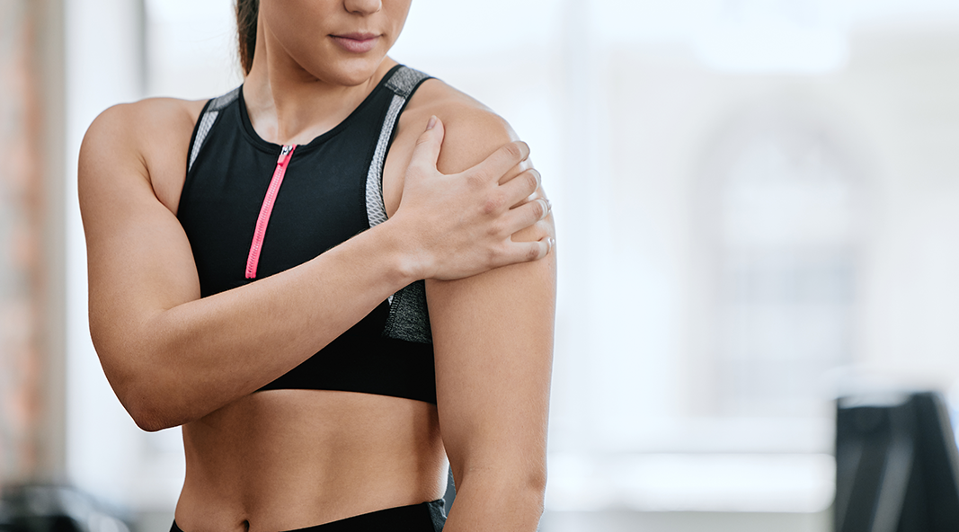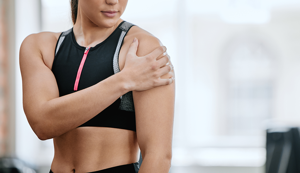
Rotator cuff injuries are common injuries among athletes. They occur when one of the four rotator cuff muscles are injured or damaged.
The rotator cuff muscles involve four muscles that surround the shoulder and are responsible for stabilizing the ball in the socket of the shoulder. They work together to provide the shoulder with its powerful range of motion, strength, and the ability to perform overhead activities.
As the muscles get closer to their insertion on the humerus, they are called tendons. There are numerous activities that may cause damage to these tendons. Sometimes the damage is small and considered “micro” from overuse and impingement. Inflammation is very common condition as well. Other times, the injury may consist of a tear, or from chronic degenerative changes of the tendon. When injuries to the rotator cuff occur, a broad spectrum of symptoms can occur and treatment for the condition will depend on the type of injury or tear, and the number of tendons involved.
In mild cases that may consist of small or partial tears, or tendonitis, Dr. Anz will usually try and treat the condition conservatively using non-operative measures first. These consist of rest, ice, anti-inflammatory medications, physical therapy, and possibly, an injection to reduce the inflammation.
In patients with full thickness tears, large partial-thickness tears, or smaller tears/tendonitis that have failed non-operative measures, surgery may be indicated.
In the majority of cases, surgery to treat rotator cuff tears can be performed arthroscopically using small incisions, a camera, and tiny instruments to perform the procedure.
During surgery, Dr. Anz will enter the shoulder joint and examine the injury by identifying the torn tendon and reattaching it to its insertion site on the humerus—this is known as the footprint, or original site of attachment. This process is completed using sutures placed through the tendon, and anchors that are attached to the bone to secure the normal anatomy of the tendon.
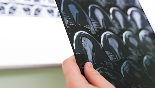
Dr. Anz will determine the exact type of repair once the configuration of the tear has been assessed.
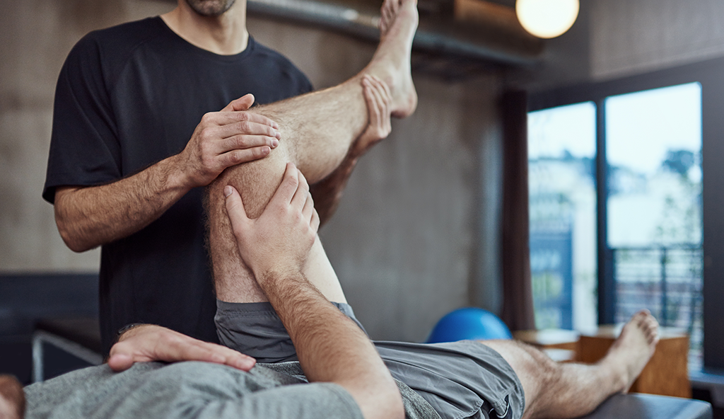
New procedures are now helpful in repairing tendons with additional support. In some cases, a double row repair will be performed if needed to provide compression of the tendon for healing. Every patient will be given a strict rehabilitation protocol to follow once surgery is completed. Immediately following post-op, patients are placed into a sling and an individualized physical therapy program is started. Rehabilitation will include the work of a skilled therapist and will involve passive range of motion, followed by active motion, strengthening, and eventually return to activities. The specific progression of therapy will depend on the configuration of the tear, type of repair, and the number of tendons involved.
—
For additional resources on rotator cuff injuries, or to learn more about arthroscopic rotator cuff repair surgery, please contact the Gulf Breeze, Florida orthopedic shoulder practice of Dr. Adam Anz located at the Andrews Institute.

