Injury Overview
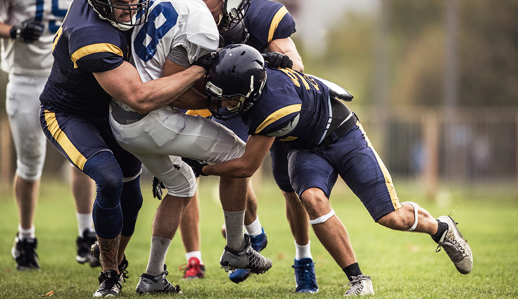
Shoulder dislocations are the result of the humeral head (ball) and glenoid area of the scapula (socket) to become pulled apart. Athletes who perform powerful overhead motions, such as serving in tennis or pitching in baseball, put the shoulder joint at risk for dislocations. While the shoulder joint offers the greatest range of motion of any joint in the human body, it also offers an extreme mobility that comes at the expense of stability.
To allow for such great motion, the shoulder is stabilized by soft tissue restraints (such as ligaments and cartilage that surrounds the socket called the labrum). When a shoulder dislocation occurs, the ball comes out of the socket and in most cases, the soft tissue stabilizers are damaged as well.
While the initial treatment for a shoulder dislocation is to reduce the joint (put the ball back into the socket), ongoing instability most likely will occur if further assessment is not performed on the joint to ensure that additional damage doesn’t exist. Once the shoulder is reduced, X-rays will then be utilized to assess any nearby damage to other bones. Dr. Anz will also determine if the patient should be treated surgically or non-surgically in order to stabilize the joint. If he feels a surgery needs to be performed, he will perform arthroscopic stabilization surgery for shoulder instability.
In the majority of patients, arthroscopic stabilization has been shown to be highly effective in eliminating shoulder instability. In certain situations such as longstanding instability, bone loss from the glenoid or humerus, and a dislocation that can’t be manually reduced, a specialized open procedure may be necessary.
During arthroscopic stabilization surgery, the shoulder is examined to confirm the direction and degree of instability. Next, the area of damage will be assessed and small surgical instruments used to place anchors into the bone on the glenoid that contain strong sutures. These sutures are then used to repair the torn labrum and ligaments, restore the anatomy of the joint to its natural position, and to effectively “tighten” the shoulder back to normal.
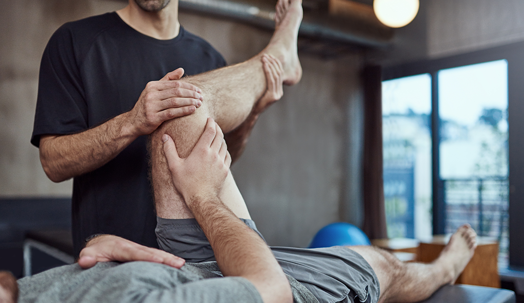
Dr. Anz will require all patients to become involved in a post-operative therapy program following arthroscopic stabilization shoulder surgery. This typically consists of gentle passive range of motion movements, followed by active motion, strengthening, and eventually, the return to activities. The patient will continue to wear a cling for about 6 weeks. Dr. Anz will examine the joint to assess how the progression of therapy should continue. This will depend on the configuration of the injury and type of repair.
For more information on arthroscopic stabilization shoulder surgery, or to learn more about your specific shoulder injury and shoulder pain, please contact the Gulf Breeze, Florida office of orthopedic shoulder surgeon, Dr. Adam Anz located at the Andrews Institute.


