Injury Overview
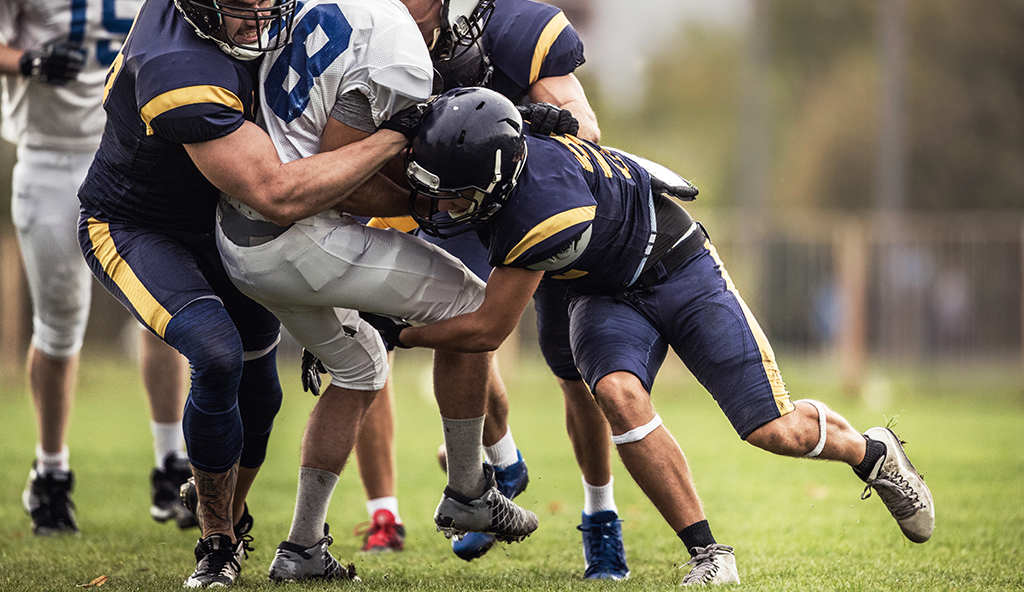
The collarbone (clavicle) of the shoulder can become fractured from a direct hit or a fall onto the shoulder. This is a common injury for athletes and frequently occurs in football players. Clavicle fractures can occur anywhere along the bone and in numerous configurations. They also can result in multiple bone fragments which is a scenario referred to as “comminution”. Most clavicle fractures can heal over time with use of a sling and avoidance of activities for 6 to 10 weeks. In comminuted fractures or fractures in which the bones do not line up well, surgery is often recommended to restore the normal alignment and length of the bone.
Clavicle fracture surgery usually involves an incision over the top of the shoulder and placement of a plate and screws along the top of the clavicle. The goal of surgery is to stabilize the clavicle so that it can heal in the appropriate position. Surgery does not speed up the healing process but rather ensures that the bone heals correctly. Certain types of clavicle fractures can also be fixed with the use of a long pin placed within the bone (intramedullary nailing). Patients are usually allowed to go home the same day as their surgery.
Recovery from clavicle fracture surgery often takes a few months and involves sling use and guided physical therapy. This included range of motion training and strengthening. Some patients request to have their hardware removed from the collarbone but this is not allowed until after the fracture has fully healed.
—
For additional information on clavicle fracture fixation surgery, or to learn more about other injuries associated with the collarbone, please contact the Gulf Breeze, Florida orthopedic surgeon, Dr. Adam Anz located at the Andrews Institute.


