Injury Overview
The gluteus muscles are a group of muscles that allow an individual to partake in rigorous activity such as running and jumping. These muscles are broad, strong muscles that make up the outer buttocks in the human body. There are two muscles to consider:
- -The gluteus medius muscle is located at the outer part of the hip.
- -The gluteus minimus is the smallest of the gluteal muscles and is located immediately beneath the gluteus medius.
Together, these muscles work to straighten the hip during activity, stabilize the pelvis and assist with outer movements of the hip. A tear in the gluteus medius muscle typically occurs at the area where the muscle attaches to the femur bone. Gluteus tears can occur from traumatic injuries which cause the tendon to peel off of the bone. However, most gluteal tears are degenerative and are caused by chronic inflammation from repetitive movements and overuse. This can sometimes be associated with trochanteric bursitis of the hip.
Symptoms
The primary symptoms of a gluteal tear include an abnormal gait, hip pain, and lower back pain. Symptoms become worse with long periods of sitting, standing, and walking. Some patients experience hip tenderness when lying on the affected side. Symptoms will also depend on the grade of the injury:
- Grade 1: Mild pain with little or no loss of mobility
- Grade 2: Partial tear with mild pain and a noticeable loss in strength and flexibility
- Grade 3: Full/complete tear; severe pain coupled with a complete loss of strength; limited mobility
Diagnostic Testing
A tear of the gluteus muscle can usually be discovered through a physical exam. Dr. Anz will conduct a series of tests to check for tenderness over the lateral hip area. Additional strength tests may reveal pain and weakness with resisted hip abduction. To rule out other injuries and conditions, Dr. Anz could order an X-ray or MRI to take a further look inside the hip and give a final diagnosis on the grade of the tear.
Treatment
Non-Surgical
For Grade 1 tears, conservative measures will be prescribed to treat the injury, such as using ice and anti-inflammatory drugs to reduce pain and inflammation. It’s recommended that patients should avoid sports or major physical activities and movement to allow healing to occur.
Surgical
In severe Grade 2 or in Grade 3 tears, Dr. Anz will repair the tear endoscopically, whereby tiny incisions are made and the torn gluteus muscle is reattached with sutures. This is minimally invasive and is achieved through the use of the camera. Repair of the tendon back to its attachment site on the greater trochanter allows for healing and restoration of function. The body will mend the torn tendon edge over a period of many weeks.
Post-Op
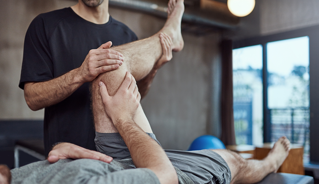
Dr. Anz will order a complete physical therapy plan after surgery. Rehabilitation of a gluteus tear focuses on gentle hip range of motion and progressive strengthening exercises, with an emphasis on hip abductor, extensor, and internal rotator muscles. Balance exercises will be introduced as strength returns.
—
For more information on gluteus medius and gluteus minimus tears of the hip, or for questions on arthroscopic hip surgery, please contact the Gulf Breeze, Florida orthopedic surgeon, Dr. Adam Anz located at the Andrews Institute.


