Injury Overview
The knee joint is a crucial component of the lower extremity mechanical axis in the human body. In orthopedics, proper alignment of the lower extremity is essential for normal joint function, muscle development, biomechanics, and dynamic balance. If the lower extremity is improperly aligned, problems can arise in the knee joint including damage to the articular cartilage and/or meniscus because forces across the knee are not evenly balanced. An improper alignment of the lower extremity is often called a malalignment. Over time, a malalignment may cause damage to the knee joint and its ligaments and can cause various symptoms in both young and older individuals.
Some people are born with a malalignment, while others can develop malalignment due to a traumatic event or damage to structures on one side of their knee.. There are a number of problems that can arise due to a malalignment, including articular cartilage damage, meniscal damage, and a ligament injury.
There are two types of malalignment in the knee:
- VARUS: The term “bow legged” refers to a varus malalignment. This occurs when weight does not pass evenly through the knee and instead passes through the inside (or medial compartment) of the knee joint (inside). With this condition, patients are more likely to develop degeneration of this side of the knee and are at a risk of having medial meniscal tears and cartilage injury. With time, patients may also stretch out the ligaments on the outside of their knee and develop instability during walking. A varus thrust involves visible instability upon walking.
- VALGUS: The term “knock kneed” refers to a valgus malalignment. This occurs when the weight-bearing axis passes through the lateral side of the knee (outside), predisposing this side to injury and wear.
Varus and valgus alignments occur on a spectrum. Some people may have a mild amount of varus or valgus which does not cause any problems. However, extreme cases of varus and valgus involve weight transmission across the knee joint which is not balanced. This can cause unequal wear in the knee joint as one side experiences greater force than the other side.

Symptoms
Some change in the mechanical axis is normal through childhood development, however, malalignments which persist after childhood can affect one’s stance and gait (pattern of walking). Patients may report that they have been “bow-legged” or “knock-kneed” since childhood. These patients may or may not develop problems later in life depending on the extent of their alignment. Patients that develop a malalignment due to other causes often report an initial injury many years ago. They then noticed a gradual onset of pain on one side of their knee. Mechanical symptoms such as knee swelling, popping, catching, and a reduced range of motion may also be present.
Diagnosis
The diagnosis of malalignment will require a physical examination as well as X-rays that capture the entire lower extremity mechanical axis. Dr. Anz will examine the hip, knee and entire lower extremities. Full length standing X-rays will document the weight-bearing axis (overall alignment) of the leg and evaluate the overall status of the knee. For patients with mechanical symptoms, an MRI will help to evaluate the cartilage and meniscus of the knee joint.
Treatment
Some patients who are diagnosed with a malalignment can be treated conservatively without surgery. A thorough understanding of the problem and avoiding certain activities can be helpful. Additionally, stretching and strengthening of the quadriceps, hamstrings, and calf muscles will help provide stability to the knee joint. Weight loss, core and lower extremity strengthening, shoe modifications, and bracing to shift the mechanical axis may also be recommended.
Surgical Treatment
In certain instances, surgery may be recommended. Surgery to correct a malalignment requires an osteotomy (or cut in a bone) and realignment. This may be performed on the leg bone (tibia) or thighbone (femur) and may also be combined with other procedures. In certain instances of a ligament injury in the setting of malalignment, a staged surgery may be necessary. For instance in patients with malalignment and chronic ligament injuries, two surgeries may be necessary. The first surgery will involve an osteotomy correction of the malalignment followed at a later date by ligament reconstruction surgery. There are also procedures to treat chondral injuries that may result from the malalignment.
Post-Operative
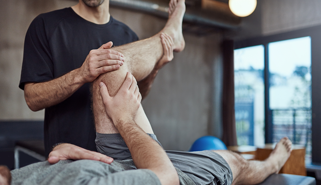
Depending on the specifics of the surgery patients may be immobilized following surgery for a period of time and may be restricted from putting weight on their leg. Physical therapy is always important and will focus on patient mobility, returning motion at the appropriate time, and regaining strength back to the injured knee and surrounding muscles. Rehabilitation is a crucial part of the recovery process and is recommended to achieve optimal results.
—
For additional resources on knee conditions involving a malalignment of the knee, such as a varus or vargus knee disorder, please contact the Gulf Breeze, Florida orthopedic surgeon, Dr. Adam Anz located at the Andrews Institute.


