Ski Knee Pain

Revision ACL Reconstruction

Ski Knee Pain

Injury
The anterior cruciate ligament (ACL) is a ligament located inside the knee and responsible for providing stability to the knee with rotational movements or twisting. For a complete discussion on injury to this ligament, see our blog article (https://adamanzmd.com/acl-knee-injuries/).   While ACL surgery is most often successful, there are occasions where reinjury can occur. In some instances, reinjury may involve a tear of a previous ACL reconstruction or repair.

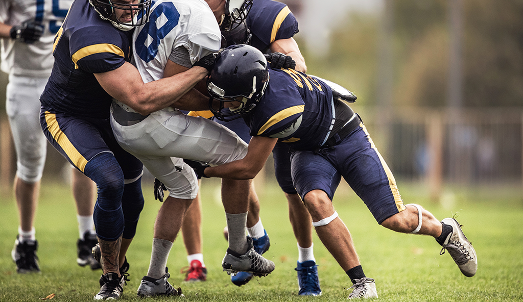
When looking at reinjury rates in the general population, one study found a 4.2% rate of revision reconstruction at 5 years1 and that patients who were young at the time of the reconstruction were more at risk of reinjury.  Another study in patients under 20 years of age who had an ACL reconstruction found that 18% reinjured either their reconstructed knee or their other knee2.   Of reinjuries, 90% occurred during high-risk sports.3  A systematic review comparing autograft and allograft reconstructions in young patients found a higher reinjury rate in patients under the age of 25 who had an allograft.  (9% and 25%, respectively).4  It is clear that reconstructions involving younger patients are more at risk of reinjury, and allografts in young patients are at a higher risk of reinjury.

Symptoms

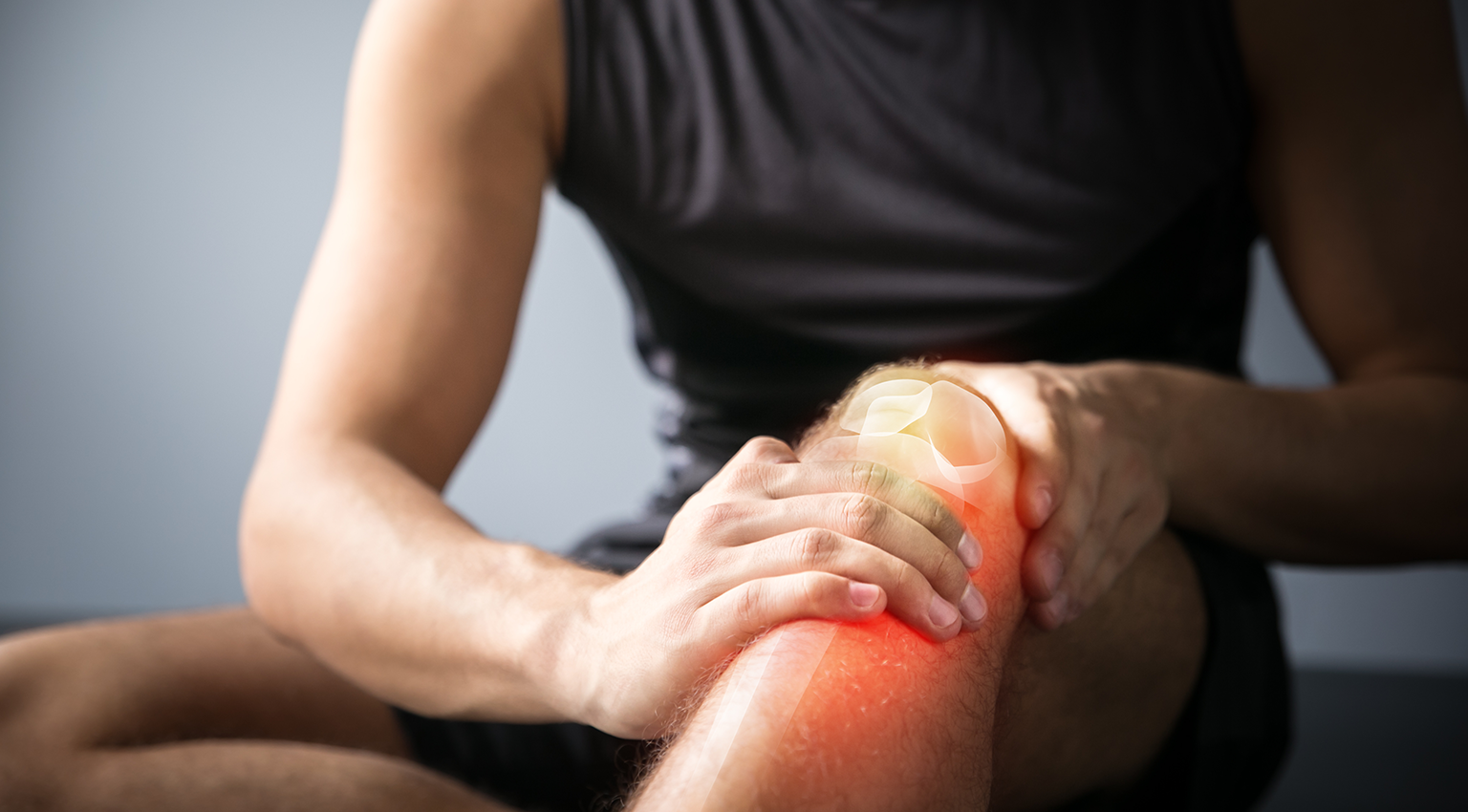
When an individual retears their ACL, there may be less pain and swelling then when they injured their knee the first time.  Patients typically report similar feelings of instability than what they felt prior to their first surgery.  In some instances, they may sustain a meniscus tear at the time of their reinjury, and the knee may be locked from full extension (straightening).  In younger, active patients a revision ACL reconstruction surgery is often recommended.

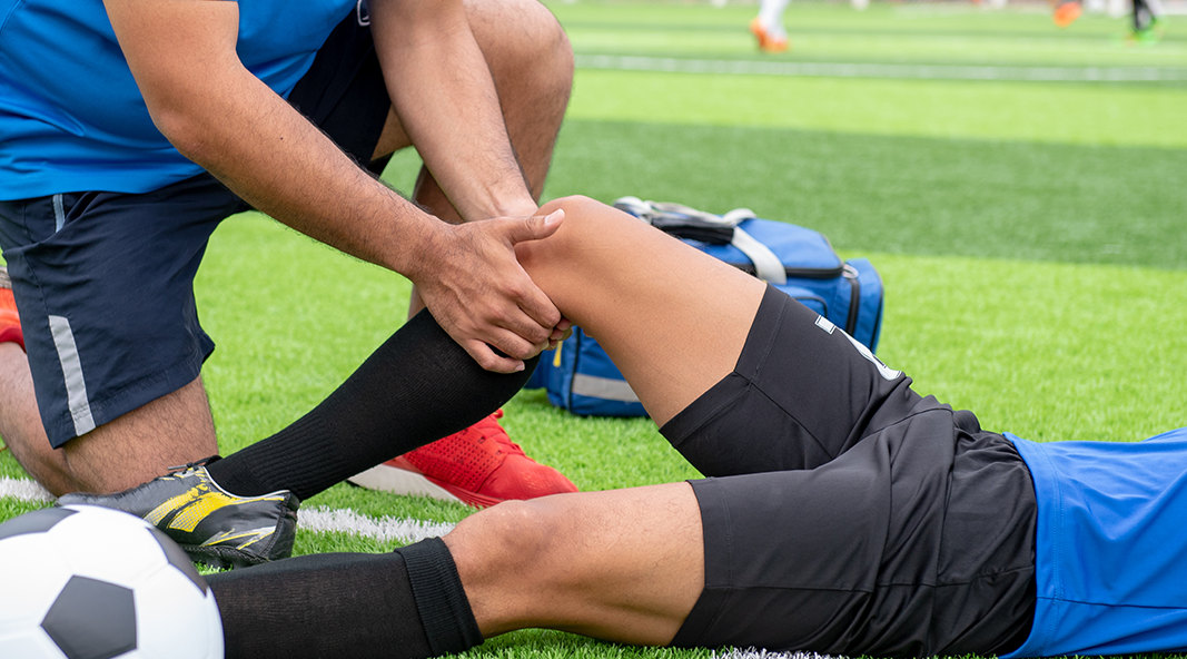
It is hard to know for certain why a re injury occurs. Some reasons may include: a too soon return to cutting/pivoting activities, too little rehabilitation, or new trauma to the knee (such as a fall or impact to the knee during sporting activity). Once a re-tear occurs, the knee is likely unstable and must be carefully addressed to restore function to the knee.
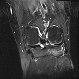
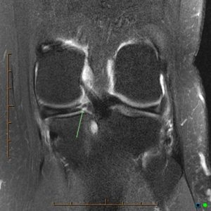
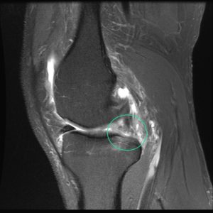
Diagnosis

Dr. Anz will assess the knee carefully and will order new X-rays and most likely an MRI.  New X-rays and MRI help to fully understand the extent of, possible reasons/risks for, and the best steps to take to address the reinjury.  In some instances, a CT scan helps to see the status of femur and tibia bones, considering the previous bone tunnels/sockets used during the first surgery.

Treatment
An ACL revision surgery may be the best way to return athletes to the level of sport which they seek.  Revision surgery is more difficult to perform because previous devices used with the first ACL surgery and the tunnels created for the first surgery affect the revision surgery.  In certain cases, a revision ACL reconstruction can be performed immediately, in one stage.  In other cases where the previous bone tunnels create hurdles, a revision surgery in two stages may be the best course of action.   In two-staged revisions,  a bone grafting surgery to fill the areas with new bone is the first stage, and a second-stage surgery , 3-6 months after the first stage, to place the new ACL reconstruction graft follows.

Post-Operative

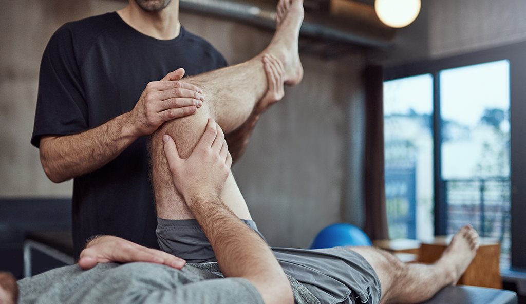
Patients will be prescribed a clear and thorough rehabilitation program following revision ACL surgery. After surgery patients will be placed into a brace and will typically use crutches for 2 weeks.  Rehabilitation will be a progressive process that may initially limit movement.  The first phase focuses on swelling and the return of normal knee motion.  Further phases focus on regaining strength, balance, and functional movement patterns.

Recovery Time

After a first ACL reconstruction, cutting and pivoting activities are limited until around the 7 month time point as graft maturation takes time.  With revision reconstructions, this may be pushed to the 9 month time point.  Two studies on reinjury rates suggest that risk decreases significantly with every month until the 9-month time point. For this reason, in most instances a return to cutting/pivoting sports is cautioned before the 9-month milestone.5  However, every athlete is unique and situations are unique.  Some instances dictate a sooner return to sport than 9-months understanding the risk.  Post-operative rehabilitation and returning to sport is a joint effort/decision between the patient, Dr. Anz, the physical therapist, and the athletic trainer and is necessary to achieve the most optimal outcome.

Athletes with ACL injuries should not feel bad if it takes time to return to their sport.  In many instances it takes athletes one to two years to make a full return.  A study in high-school and college football players found a return to sport rate of 32% at one year and 64% at two years.  53% of high school players and 50% of collegiate players identified fear as a major or contributing factor to not returning to play sooner.6 Dr. Anz and his team serve as advocates to help athletes return safely and expeditiously to their sport, considering and helping with all hurdles along the way.

For additional information on revision ACL reconstruction surgery, or to learn more about common knee injuries involving one or more ligaments within the knee, please contact the Gulf Breeze, Florida orthopedic surgeon, Dr. Adam Anz located at the Andrews Institute.

Sources: 

  1. PubMed.gov, Authors: Andreas PerssonKnut FjeldsgaardJan-Erik GjertsenAsle B KjellsenLars EngebretsenRandi M HoleJonas M Fevang. Date: Dec 9, 2013. Link.
  2. PubMed.gov, Authors: Sue Barber-WestinFrank R Noyes. Date: May 6, 2020. Link.
  3. PubMed.gov, Authors: Hideaki FukudaTakahiro OguraShigehiro AsaiToru OmodaniTatsuya TakahashiIchiro YamauraHiroki SakaiChikara SaitoAkihiro TsuchiyaKenji Takahashi. Date: Dec 9, 2013. Link.
  4. PubMed.gov, Authors: David WassersteinUjash ShethAlison CabreraKurt P Spindler. Date: May 7, 2015. Link.
  5. PubMed.gov, Authors:
    Susanne Beischer, PT, PhD, Linnéa Gustavsson, Eric Hamrin Senorski, PT, PhD, Jón Karlsson, MD, PhD, Christoffer Thomeé, BS, Kristian Samuelsson, MD, PhD, Roland Thomeé, PT, PhD. Date: Jan 31, 2020. Link.
  6. PubMed.gov, Authors: Kirk A McCulloughKevin D PhelpsKurt P SpindlerMatthew J MatavaWarren R DunnRichard D ParkerMOON GroupEmily K Reinke. Aug 24, 2012. Link.
Biking Knee Pain

When Is An Athlete Ready For Contact Sports After ACL Surgery?

Biking Knee Pain

Returning to Sport Following ACL Reconstruction

After ACL reconstruction1, the most common question is also the most difficult to answer: “When will I be ready to go back to ___?” In short, there is no blanket answer; there are many factors that determine when the time is right for a patient to return to sport. These factors are physical, biologic, and psychological in nature and they affect each patient’s recovery in a unique way. At the end of the day, returning to sport is a decision that needs to be made on a patient-to-patient basis, weighing the benefits of continued rest and rehabilitation with the risks and benefits of returning to sport.

How and When Do Most Athletes Return

It is helpful to consider the track record of other athletes’ road to recovery following ACL reconstruction. Traditional wisdom holds that athletes return to sport within 6-12 months of an ACL injury. However, recent studies have turned this idea on its head. One study evaluated 187 amateur and competitive athletes with ACL injuries and found that only 31% returned to the sport in the first 12 months, and that number only climbed to 60% at 24-months post-surgery.2 Another multicenter study examined the question with a specific focus on football. Researchers contacted 147 high school and collegiate football players and found similar return-to-play rates in both groups two years post-surgery—63% and 69%, respectively. However, only 43% of the players interviewed believed they returned to previous self-reported levels of performance; 27% felt that they never reached their pre-surgery performance and 30% were unable to return at all. Surprisingly, fear of re-injury or further damage to the knee was cited as the most common reason that players did not return to play.3 This study highlights two important facts: 1) return-to-play rates for football players are not as high as one might expect, and 2) psychological factors, particularly fear of re-injury, play a key role in athletes’ return to sport.

Physical Therapy: Motion, Muscle Strength, and Proprioception

Physical therapy following surgery is just as, if not more, important than the surgery itself. After most ACL reconstructions, full restoration of range of motion in the knee is the first goal of therapy, except in cases involving complex meniscus repairs. Once motion is restored, strength becomes the next goal. During this phase, athletes may find that it takes longer than expected to recover pre-injury strength and mass in the muscles surrounding the knee. This can be frustrating, and it is surprising how different patients recover at different rates. The key during the strengthening phase is to work hard and be patient. While strengthening the muscles, therapists will also help restore normal/athletic proprioception and functional movement patterns. At this point, it is common for athletes to not experience pain, yet still have significant strength and coordination deficits.

A physical therapist is your most important resource during rehabilitation. They have the experience and skills necessary to help athletes realize where they are in the process, set appropriate goals, and find a balance between activity and rest—both of which are critical to recovery. The athlete, their family, their therapist, athletic trainers, and the physician are all part of a team working together during the recovery process. This process takes time—anywhere from 6 to 24 months—and can vary due to the extent of injury, the athlete’s stage of life/career, and how quickly one’s body recovers. The key is to be patient and persistent, allowing the entire team to help guide the process.

“No Doubt”- The Return of the Confident, Athletic Mindset

Confidence and risk are the final factors that must be considered as an athlete prepares to return to sport. As highlighted earlier, 50% of athletes cite fear of reinjury as the primary obstacle to returning to competition. There are two potential explanations for this fear: 1) a graft that “just doesn’t feel right” may not have completed the ligamentization process, or 2) traditional rehabilitation does not focus on the athlete’s competitive mindset.

While research seeks to eliminate the first cause—and our next blog will address these efforts in detail—only a team-based approach, consisting of the athlete and their family, the physical therapist, athletic trainers, and physician can resolve the second. Close communication is important to understand risks and expectations, set and achieve rehabilitation goals, and confirm the return of normal function and strength. While it is ultimately up to the athlete and their family to decide when he or she is prepared to return to full competition, at the Andrews Institute in Gulf Breeze, Florida, Dr. Anz, and his team work together to aid you in this difficult time.

“The Sports Test-” Progress Report of Recovery

Dr. Anz’s team uses a ‘Sports Test’ to determine where an athlete is in the rehabilitation process. The test is usually scheduled at the eight- to nine-month mark, depending on the athlete’s goal for return to sport. The test evaluates strength through a mechanical test called a Biodex test, and coordination, proprioception and balance through functional movement screening and a y-balance test. There are many versions of this test, but their main goal is to objectively grade an athlete’s strength, coordination, proprioception, and functional movement patterns and, therefore, determine when they are physically equipped to return to sport. Again, there is no standard timeframe outlining when an athlete can return to competition. Therefore, Dr. Anz works with his medical team to evaluate each athlete and determine the goals they have met and the goals that still need to be accomplished. It is not uncommon for previously unknown deficits to be uncovered during a Sports Test. Through this identification, the athlete’s rehabilitation team can refocus and continue their efforts.

The Andrews SCORE: An Evidence-Based Multi-model Return to Sport Evaluation

At the Andrews Institute, a project is starting in 2022 to develop a data-driven decision making model to assess a competitive athlete’s readiness to return safely to sport after ACL injury and surgery.  The model will include patient-reported outcome measures, objective and modern strength/ability testing, and MRI evaluation of healing tissues.  The MRI evaluation will involve techniques already studied by the Andrews Institute and Auburn University.4  This return to sport algorithm will target competitive athletes with a return to sport that is congruent with their pre-injury level of play.  The goal is to have a system to confirm that injured athletes are ready physically to return safely to their sport.  This system will then allow for further study of biologics to expedite this return, proving readiness before return. The ligamentization process of the graft is always the wild card of knowing when athletes are ready for a full return.5  Our goal is to study if further and advance the biologic capabilities of ACL surgery.


It is critical to always keep the athlete’s best interests in mind, understanding the short-term gain and long-term health consequences of every decision.

As an Orthopedic Surgeon and Sports Medicine Specialist, Dr. Adam W. Anz is dedicated to providing individuals and athletes from all over the world with the highest possible quality of care. He serves his patients at the world-class Andrews Institute in Gulf Breeze, Florida. If you have sustained a sports-related injury, please contact our office today to schedule an initial consultation with Dr. Anz.

Sources:

  1. https://adamanzmd.com/acl-reconstruction/
  2. https://journals.sagepub.com/doi/abs/10.1177/0363546514563282?journalCode=ajsb
  3. https://pubmed.ncbi.nlm.nih.gov/22922520/
  4. https://adamanzmd.com/wp-content/uploads/2021/11/2019-3T-MRI-mapping-is-a-valid-in-vivo-method-of-quantitatively-evaluating-the-anterior-cruciate-ligament-rater-reliability-and-comparison-across-age.pdf
  5. https://adamanzmd.com/thebiologyofACLhealing-thewildcardofrecovery/
Family Hiking.

ACL Knee Injuries

Family Hiking.

Injury Overview

ACL knee injuries are among the most common injuries for athletes.  The ACL (anterior cruciate ligament) is one of four ligaments that make up the knee joint and is responsible for keeping the shinbone (tibia) from sliding forward on the thigh bone (femur) and providing stability for movements requiring rotation of the knee. Cutting and pivoting athletes, including soccer players, football players, basketball players, and skiers, are at a higher risk for developing an ACL knee injury because of the sudden pivots, twists, and turns associated with these sports. The ACL can also be torn in cases that do not involve sports, such as tripping, missing a step, or any other traumatic hit to the knee.  Typically, the knee sustains a “pivot shift” event where the bones of the knee shift in an abnormal way.

Symptoms

Patients who have an ACL injury are typically present after a twisting injury event. If during a practice or competition, athletes typically report that they could not continue to perform/compete during that day and that the knee swelled immediately after the injury.  Swelling inside the knee from the injury routinely causes it to swell to the size of a softball.  Sometimes, athletes report the sound of one or more pops.  

After a few days, the swelling improves and the function returns. After an ACL injury,  recurrent feelings of “giving away” or instability of the knee are common. These episodes can be problematic because continued pivot shifting events can be associated with further injury to the knee, including injuries to the medial meniscus.   The knee can shift with certain movements such as pivoting to open a door or cutting with an attempt to return to sport.

Diagnosis

Listening carefully to the athlete’s history of injury is always the most important first step to knowing the problem.  Examining the patient thoroughly is the second step, and the Lachman test, anterior drawer test, and pivot shift test are ways to test the ACL for injury.  These tests are not painful; however, it is understandable to be apprehensive of tests of knee stability.  X-rays are important to see the bone anatomy of an injured athlete, as overall alignment affects the forces on ligaments and knees, and to rule out fractures.   MRI is an additional necessary test when an ACL is expected in order to confirm the diagnosis and look for associated injuries, as in many instances the lateral meniscus is injured at the time of an ACL injury.

Treatment

Nonoperative Treatment

Depending on the patient’s lifestyle and goals, the ultimate objective is to return the patient to their pre-injury activities.   This involves an individualized treatment plan to regain stability and full mobility of the knee.  In some cases of isolated ACL injury, surgery may not be necessary, depending on a patient’s goals. Conservative treatment consists of rest, ice, elevation, anti-inflammatory medications, and physical therapy to strengthen the muscles around the knee and to improve neuromuscular control around the knee.  An ACL brace can also help protect the joint during the rehabilitation process.

Surgical Treatment

In athletes who are returning to cutting and pivoting activities, ACL reconstruction surgery is most often recommended.  In some instances, an ACL repair can be considered. During ACL reconstruction surgery a tendon graft of a similar size is used to replace the injured tissue. Reconstruction surgery is performed arthroscopically and involves removing the injured tissue and creating a new ligament with a graft. The graft used during the reconstructive process is most often taken from the patient.   The central third of the patellar tendon, the central third of the quadriceps tendon, or two of the four hamstring tendons are the most commonly used grafts.   Although donated tissue using a graft bank from a donor can also be used, it has been associated with a higher reinjury rate in some studies.  Graft choice is individually decided with the patient.

Post Operative

After a reconstruction surgery, a thorough physical therapy program is extremely important for rehabilitation.  Rehabilitation will be a progressive process that may initially limit movement.  The first phases focus on swelling and the return of normal knee motion.  Further phases focus on regaining strength, balance, and functional movement patterns.

Recovery Time

After an ACL reconstruction, cutting and pivoting activities are limited until around the 7 month time point as graft maturation takes time.  Two studies on reinjury rates suggest that risk decreases significantly with every month until the 9-month time point.1,2  

For this reason, in most instances a return to cutting/pivoting sports is cautioned before the 9-month milestone.  However, every athlete is unique and situations are unique.  Some instances dictate a sooner return to sport than 9-months understanding the risk.  Post-operative rehabilitation and returning to sport is a joint effort/decision between the patient, Dr. Anz, the physical therapist, and the athletic trainer and is necessary to achieve the most optimal outcome.

Athletes with ACL injuries should not feel bad if it takes time to return to their sport.  In many instances it takes athletes one to two years to make a full return.  A study in high-school and college football players found a return to sport rate of 32% at one year and 64% at two years.  53% of high school players and 50% of collegiate players identified fear as a major or contributing factor to not returning to play sooner.3 Dr. Anz and his team serve as advocates to help athletes return safely and expeditiously to their sport, considering and helping with all hurdles along the way.

References:

For additional information on ACL knee injuries, or to learn more about what is involved during ACL reconstruction surgery, please contact the office of orthopedic knee surgeon, Dr. Adam Anz, serving the greater Pensacola, Gulf Breeze, Florida communities.

Media:

ACL Injury Healing

The Biology of ACL Healing: The Wild Card of Recovery

ACL Injury Healing
Ligamentization – The Wild Card After an ACL injury1, reconstruction of the ligament is often a recommended treatment.  One of the most critical steps in ACL reconstruction2 is grafting a tendon into the knee to replace the damaged ACL. Following surgery, the graft not only has to heal tightly in its new position but also transition structurally from a functioning tendon to a functioning ligament—a process known as ligamentization. Believe it or not, this process takes time! Microscopic studies, in which physicians have taken small samples of healing ligaments following ACL reconstruction to determine their rate of ligamentization, suggest that this process can take anywhere from 6 to 24 months. The ligamentization process is frustrating because it is an unseen rate-limiting step in an athlete’s recovery. Some athletes regain motion, strength, and proprioception at the six-month mark and appear outwardly healed, but the level of ligamentization of their graft is unknown and therefore a return to sport can jeopardize their recovery. For this reason, it is ideal to allow athletes recovering from ACL reconstruction as much time for rehabilitation as logistically possible, assuming it doesn’t jeopardize their return career goals.  Two studies on reinjury rates suggest that risk decreases significantly with every month until the 9-month time point.3,4  This time delay is directly related to the ligamentization process.  For this reason, in most instances, a return to cutting/pivoting sports is cautioned before the 9-month milestone.  Many instances of ACL reconstruction failure can be attributed to a failure of graft incorporation and/or the ligamentization process, so giving the athlete adequate time for recovery is best. One of Dr. Anz’s most passionate clinical research interests revolves around using biologics to improve and/or expedite the ligamentization process following ACL reconstruction. His interest is driven by how critical the ligamentization process is to the full recovery and return to the sport of injured athletes. Animal studies have suggested that slower graft incorporation correlates with increased laxity and stiffness, increasing the likelihood of re-injury, and have illustrated improved tendon healing in ACL reconstructions that incorporate the use of stem cell technologies.  We theorize that optimization of stem cell technologies for tissue regeneration requires the use of the ‘regenerative triad’—a scaffold, stem cells, and growth factors. For that reason, regenerative models with ACL reconstruction combine biologic technologies with a scaffold wrap to produce a new “sheet” of cells around the ACL graft. There are two studies that illustrate the effectiveness of biologics in improving the rate of ACL maturation. The first of these studies involved injecting leukocyte-poor platelet-rich plasma into the fascicles of ACL grafts and the other involved loading of a gelatin carrier with platelet-derived growth factors. Prior to the injury, the ACL and PCL are covered by a synovial lining—essentially a layer of collagen that ensures adequate blood and nutrient supply to the ACL. It is now believed that the lack of this synovial lining following traditional ACL reconstruction may delay the process of ligamentization. We believe that the use of a collagen membrane will protect and create a healing environment like the synovial lining of a healthy joint. At the Andrews Institute, we are interested in building a scientific body of work “brick by brick” to advance the biology of ligamentization.  In 2016, we completed a study determining how many cells can be collected from a patient’s knee, including swelling and by-products of the reconstruction surgery at the time of ACL surgery.5 This study is helping us to better understand ideal methods for collecting a patient’s stem cells.  In 2019, Dr. Anz and colleagues at Auburn University completed a study using MRI to map the ACL to develop normative values for future ACL maturation studies.6   In 2020, we completed a study that used blood concentrating devices (PRP machines) to harvest stem cells from the injury fluid at the time of ACL reconstruction for use at the time of the surgery.7  In 2021, Drs Anz, Jordan, Ostrander, and Andrews completed a study on biologic augmentation of traditional ACL reconstruction.  The theory was that collagen membranes can be used to re-establish the natural synovial lining of the ACL and that the collagen would serve as a container to hold biologic adjuncts around the ACL. This study combined collagen-wrapped graft tissue with cells from the patient’s bone marrow fluid.  This study found that wrapping a graft with an amnion collagen matrix and injecting bone marrow aspirate concentrate was associated with lower MRI T2* values, a surrogate measure of improved graft maturation and collagen content. While early studies are promising, further development is needed to determine if the ligamentization process can be sped up.  At this time, no change in rehab protocols or strong recommendations can be recommended, but the future looks bright.  It is important at this time, even with biologic adjuncts available, to stick to 9-month return to sport timelines.  There is a significant risk of reinjury if athletes push too fast too soon based on exciting theories.  With more time and study, evidence will emerge as to whether biologics can improve ligamentization.  In the meantime, we continue to work on the science to find the truth- “brick by brick”. As an Orthopedic Surgeon and Sports Medicine Specialist, Dr. Adam W. Anz is dedicated to providing individuals and athletes from all over the world with the highest possible quality of care. He serves his patients at the world-class Andrews Institute in Gulf Breeze, Florida. Sources: 1.) https://adamanzmd.com/acl-knee-injuries/ 2.) https://adamanzmd.com/acl-reconstruction/ 3.) https://www.ncbi.nlm.nih.gov/pmc/articles/PMC4912389/pdf/nihms782171.pdf 4.) https://www.jospt.org/doi/full/10.2519/jospt.2020.9071 5.) https://adamanzmd.com/wp-content/uploads/2021/11/2017-Viable-Stem-Cells-Are-in-the-Injury-Effusion-Fluid-and-Arthroscopic-Byproducts-From-Knee-Cruciate-Ligament-Surgery.pdf 6.) https://adamanzmd.com/wp-content/uploads/2021/11/2019-3T-MRI-mapping-is-a-valid-in-vivo-method-of-quantitatively-evaluating-the-anterior-cruciate-ligament-rater-reliability-and-comparison-across-age.pdf 7.) http://box5216.temp.domains/~adamanzm/wp-content/uploads/2021/11/2021-Synovial-Stem-Cells-Harvested-with-PRP-Device.pdf   Additional ACL blog articles: 1. ACL Knee Injuries 2. When is an athlete ready for contact sports after ACL surgery? 3. ACL Reconstruction 4. Revision ACL Reconstruction 5.ACL Knee Injuries 6. Save the Menisci: Meniscal Root Tears 7. Meniscus Repair Paper Published in the American Journal of Sports Medicine 8. FCL Reconstruction 9. PCL Reconstruction 10. Multi-Ligament Knee Reconstruction 11. MCL Reconstruction 12. PCL Reconstruction 13. FCL/LCL Knee Injuries

Save the Menisci: Meniscal Root Tears

The menisci are c-shaped, rubber-like cartilage discs that reside inside the knee joint.  There are two menisci in every knee, one on the inner side (medial meniscus) and one on the outer side (lateral meniscus). Their function is to increase surface area for weight transmission between the cartilage on the ends of the bone, which decreases pressure between the ends of the bone and adds to stability of the joint.  Both the medial and lateral menisci have stout attachments at the front and back of the tibia, these attachments are often called “roots”. These meniscal roots are important because they hold the meniscus in place to provide stability to the entire meniscus. The stability is obtained by a functional circumferential hoop which the C-shape obtains with attaching at the roots. This functional hoop stability prevents the meniscus from extruding out when pressure is exerted across the joint, effectively keeping the meniscus in place between the two bones.

Meniscus tears can occur in a number of different shapes and scenarios. They can occur as a result of accumulative wear and tear of the joint or as the result of an injury. Sometimes, wear and tear changes in the meniscus can be subtle until an injury event occurs and the scenario is worsened drastically. Tears of the meniscus root are especially concerning because they compromise the functional hoop property of the meniscus, rendering the meniscus non-functional. When there is a tear of the meniscal root the studies have suggested that pressure upon the cartilage is increased to levels similar to having no meniscus at all. This can subsequently cause early degeneration of the joint.

Meniscus root tears are often seen in two groups of patients:

  • The first group consists of young adults and athletes who sustain a root tear with a severe knee injury. This may include injury to the ACL, PCL or other ligaments of the knee. Failure to repair the meniscal root in these circumstances can lead to the development of early osteoarthritis, failure of a ligament reconstruction graft and other potential problems with age.
  • The second group of patients are middle-aged adults. In this population, the injury is “acute on chronic”. There are degenerative changes at the root and then the meniscal root becomes non-functional with an injury event. A sudden knee bending event accompanied by a “pop” in the back of the knee, is often described by patients who have had a root tear. A sudden deep squat or twist are also sometimes described. In this group of patients, rapid development of osteoarthritis can occur.

Symptoms

The primary symptoms of a meniscus root tear include pain on the inside or outside of the knee with mechanical symptoms.  Certain activities such as pivoting, running, climbing, or even getting up from a chair may produce symptoms including popping and catching.  Patients may hear or feel a clicking sound with movement, and the knee may be tender to the touch for some in specific locations.

Figure 1, Coronal view of normal meniscal root

Diagnosis

While an X-ray will not show meniscal damage, it is necessary to evaluate the overall health of the knee joint, as subtle changes on X-rays are common and help to guide treatment. Although a history, physical exam and x-ray are important in diagnosis, an MRI is important to visualize the meniscal root (Figure 1). These root tears can be very difficult to identify on MRI but is most often diagnosed when a “ghost sign” is seen. Meniscal root tears can be seen on coronal, axial and sagittal MRI views. On the sagittal view, as seen in figure 3, there is a ghost sign which is indicative of a meniscal root tear. A normal, healthy meniscus should look like a dark black triangle. On the coronal view, as seen in figure 2, there is a tear of the meniscal root.

Figure 2, Coronal View with “Ghost Sign”


Figure 3, Sagittal View with “Ghost Sign”

Surgical Treatment

Treatment of meniscal root tears can be very difficult, especially in older patients. In older patients, repair can be difficult as tears are not commonly diagnosed until progression of arthritis is more severe. In younger patients, repair is much easier due to decreased prevalence of joint degeneration.

Figure 4, Normal Meniscal Root

An arthroscopic approach is utilized to repair the meniscal root.  Once access is made into the knee, Dr. Anz will visualize the meniscal root (Figure 4). A device is used to pull on the root to confirm the presence of a tear. After a tear is confirmed, Dr. Anz will use a guide to drill a tunnel at the anatomic site of the original root. (Figure 5) This tunnel will become the new home for the torn meniscal root. Sutures will be passed through the torn portion (Figure 6) of the meniscus and shuttled down into the tunnel previously drilled. Once the sutures have been pulled into the tunnel, Dr. Anz will visualize the meniscus and tighten the sutures and secure them with a suture anchor.

Figure 5, Drill bit coming up from root attachment

Figure 6, Sutures passed through the root repair

Post-Operative

After surgery, the patient will be non-weight bearing for 6 weeks to allow for healing of the repair. Physical therapy may be initiated the day after surgery. Range of motion at the knee is limited to 90 degrees of flexion for the first weeks in order to prevent excess stress on the repair. Six weeks after surgery a partial protective weight bearing program is initiated to slowly wean from the crutches. Patients should avoid impact activities, deep squats, and lifting in a deep squat for a minimum of 4 months after surgery to protect the meniscus root repair.

For more information on meniscus injuries, or to learn more about the surgical treatments for meniscus tears offered by Dr. Adam Anz, Orthopedic Surgeon/Sports Medicine Specialist, please contact our office

[social_warfare buttons=”facebook,twitter”]

Directions & Parking Information

Our office is located at:

The Andrews Institute
1040 Gulf Breeze Pkwy Suite 203
Gulf Breeze, FL 32561
Phone: 850-916-8476
FAX:  850-916-8764

[wpgmza id=”1″]


From the North

  • Travel South on I-110 to HW 98 East
  • Travel across the Pensacola Bay Bridge (aka the 3-mile Bridge) into Gulf Breeze
  • Follow Hwy 98 East for approximately 1.2 miles
  • Turn left into the driveway at the 4th spotlight (you will pass the exit to the beach and drive under an overpass). The fourth light is at the entrance to Gulf Breeze Hospital.
  • Take the firs left into the Andrews Institute
  • Turn left into parking lot

From the East

  • Travel West on Hwy 98 into Gulf Breeze
  • Go through Gulf Islands National Seashore to the stoplight (entrance to Gulf Breeze Hospital)
  • Turn right at the light
  • Take first left into the Andrews Institute
  • Turn left into parking lot

From Pensacola International Airport (PNS)

(Download Directions)

  • Travel East on Airport Blvd
  • Turn left onto North 12th Ave
  • Turn right onto East Fairfield Dr
  • Use left lane to turn onto Interstate 110 South and travel south toward beaches
  • Take exit 1B toward Gregory St/US-98/Beaches and continue onto Bayfront Pkwy
  • Continue onto East Gregory St
  • Continue onto US-98/Pensacola Bay Bridge
  • Turn left into the driveway at the 4th spotlight (you will pass the exit to the beach and drive under an overpass). The fourth light is at the entrance to Gulf Breeze Hospital.
  • Take the firs left into the Andrews Institute
  • Turn left into parking lot

From Destin-Fort Walton Beach Airport (VPS)

(Download Directions)

  • Travel West on FL-85/Eglin Pkwy
  • Turn right onto General Robert M Bond Blvd.
  • Turn slight right onto FL-189/Lewis Turner Blvd. Continue to follow FL-189.
  • Turn Right onto Mary Esther Cut Off NW/FL-393. Continue to follow FL-393.
  • Turn right (West) onto US-98 into Gulf Breeze
  • Go though Gulf Islands National Seashore to the stoplight (entrance to Gulf Breeze Hospital)
  • Turn right at the light
  • Take the first left into Andrews Institute
  • Turn left into parking lot

Parking for the Andrews Institute

  • Patients may park in front of the main entrance or Andrews Research and Education
  • Complementary valet parking is available under the overhang at the main entrance Monday-Friday from 7 a.m. to 5 p.m.
Biceps Anatomy

Biceps Anatomy Study

Biceps Anatomy

Thank you to Eric Branch for his help with our recent study on the biceps femoris. It was published last month in the Orthopaedic Journal of Sports Medicine. 

This tendon injury can accompany anterior cruciate ligament injuries in football players. The more we learn about it the better we will be at our repairs/reconstructions. In this study we learned exactly where the tendon inserts on the tibia and fibula and its association with the anterolateral ligament.

Read More:

http://www.ncbi.nlm.nih.gov/pubmed/26535398

Meniscus Repair Paper Published in The American Journal of Sports Medicine

A biomechanical study that we completed at the Andrews Research and Education Foundation was published in the American Journal of Sports Medicine.

The study evaluated the repair strengths of four meniscus repair methods for radial meniscus tears. Radial meniscus tears can accompany anterior cruciate ligament (ACL) tears, particularly radial tears of the lateral meniscus. A new instrument known as the Ceterix Novostitch was evaluated.

The study determined that complex patterns of suture repair are stronger than simple patterns.

To read the full study click here.

Lyon France Orthopedics Conference

Scientific Presentation in Lyon, France

Lyon France Orthopedics Conference

Dr. Anz presented his research on meniscus repairs at this years International Society of Arthroscopy, Knee Surgery, and Orthopaedic Sports Medicine in Lyon, France.

The study evaluated a new method to repair radial meniscus tears with a new instrument, the Ceterix Novostitch.