Loose Bodies Orthopedic Surgery

Loose Bodies

Loose Bodies Orthopedic Surgery

Injury Overview

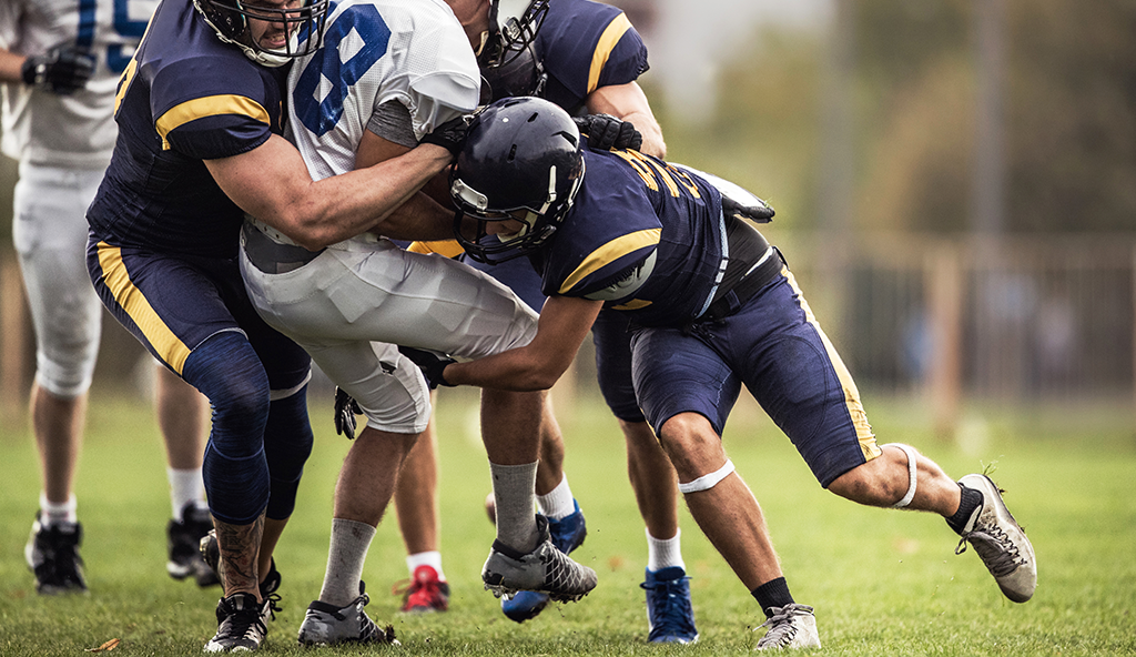
Sometimes structures can become dislodged and mobile inside a joint.  When this occurs these structures are called loose bodies.  Loose bodies can be made of bone, cartilage, or items which do not normally belong in a joint.  They can arise from an injury or from the wear and tear changes which joints go through with time.  For some patients, loose bodies within a joint do not cause pain or disability.  They can go unnoticed for years.  However, for some people loose bodies can cause significant problems.  Some individuals with loose bodies in a joint will find that with certain movements mechanical symptoms, such as popping, catching or locking, will occur. This is may be caused by these loose fragments moving to different regions or becoming lodged between two structures in a joint.  In a hip, loose bodies can be a source of significant discomfort and pain.  When loose bodies are determined to be the cause of pain, they can be surgically removed.  Without treatment symptoms may continue to worsen and healthy articular cartilage may become further damaged contributing to further joint degeneration.

Symptoms

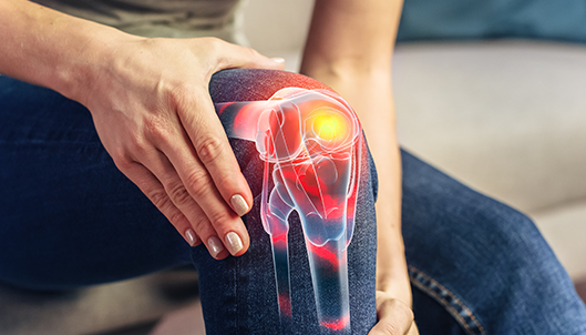
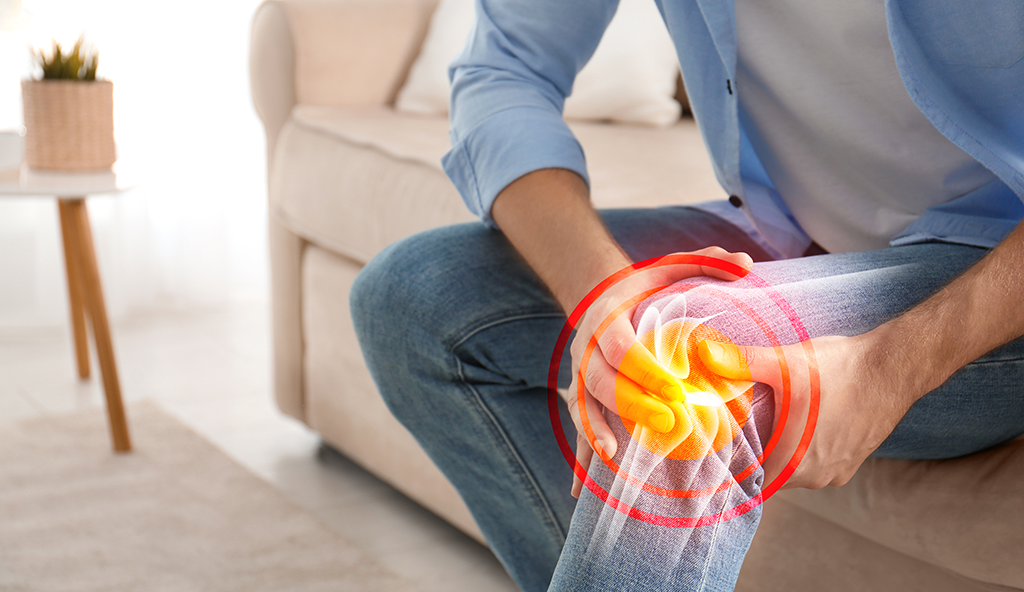
When loose bodies within a joint do cause symptoms, these symptoms often occur after reproducible specific movements.  Patients often begin to recognize pain with certain activities or motions and begin to alter their movement or activity.  The most common symptoms associated with loose bodies are:

  • Sharp pain during a specific motion
  • A feeling of “catching” inside the hip
  • A feeling of “locking” inside of the hip
  • Sensations of instability
  • A constant dull ache

Diagnosis

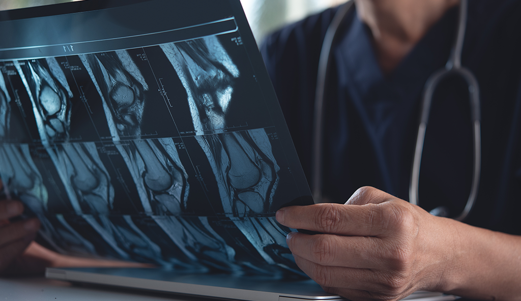
Loose bodies often arise from a specific injury. For this reason, Dr. Anz will get a full patient history to understand any previous injuries which may have occurred.  He will follow this with a physical examination and evaluation of X-rays and potentially a MRI to determine where the loose bodies reside and if there are any additional injuries to assess.  Typically, the only treatment for symptomatic loose bodies is surgical removal.

Surgical Removal

Dr. Anz prefers an arthroscopic surgical approach to remove loose bodies from the hip joint. It is not always possible to remove all loose bodies depending on a given scenario.  During arthroscopic hip surgery, keyhole incisions are made around the hip through which a small camera and small surgical instruments are used. The camera allows for visual location of loose fragments of cartilage and bone.   After visualization, grasping instruments may be used to remove these fragments.  During this process he may also use additional tools to smooth and reshape rough areas that have suffered damage due to the loose bodies.

Post-Operative

Following arthroscopic hip surgery, Dr. Anz will prescribe a progressive rehabilitation plan whereas the patient will work closely with a therapist to gain range of motion, strength, and movement. Typically, patients are able to resume their normal activities within 6-8 weeks following surgery.

If you have any questions regarding hip pain or arthroscopic hip surgery, or would like additional information on loose bodies, please contact the Gulf Breeze, Florida orthopedic surgeon, Dr. Adam Anz located at the Andrews Institute. 

Gluteus Medius Minimus Tears

Gluteus Medius/Minimus Tears

Gluteus Medius Minimus Tears

Injury Overview

The gluteus muscles are a group of muscles that allow an individual to partake in rigorous activity such as running and jumping. These muscles are broad, strong muscles that make up the outer buttocks in the human body. There are two muscles to consider:

  • The gluteus medius muscle is located at the outer part of the hip.
  • The gluteus minimus is the smallest of the gluteal muscles and is located immediately beneath the gluteus medius.

Together, these muscles work to straighten the hip during activity, stabilize the pelvis and assist with outer movements of the hip. A tear in the gluteus medius muscle typically occurs at the area where the muscle attaches to the femur bone. Gluteus tears can occur from traumatic injuries which cause the tendon to peel off of the bone. However, most gluteal tears are degenerative and are caused by chronic inflammation from repetitive movements and overuse. This can sometimes be associated with trochanteric bursitis of the hip.

Symptoms

The primary symptoms of a gluteal tear include an abnormal gait, hip pain, and lower back pain. Symptoms become worse with long periods of sitting, standing, and walking. Some patients experience hip tenderness when lying on the affected side. Symptoms will also depend on the grade of the injury:

  • Grade 1: Mild pain with little or no loss of mobility
  • Grade 2: Partial tear with mild pain and a noticeable loss in strength and flexibility
  • Grade 3: Full/complete tear; severe pain coupled with a complete loss of strength; limited mobility

Diagnostic Testing

A tear of the gluteus muscle can usually be discovered through a physical exam. Dr. Anz will conduct a series of tests to check for tenderness over the lateral hip area. Additional strength tests may reveal pain and weakness with resisted hip abduction. To rule out other injuries and conditions, Dr. Anz could order an X-ray or MRI to take a further look inside the hip and give a final diagnosis on the grade of the tear.

Treatment

Non-Surgical

For Grade 1 tears, conservative measures will be prescribed to treat the injury, such as using ice and anti-inflammatory drugs to reduce pain and inflammation. It’s recommended that patients should avoid sports or major physical activities and movement to allow healing to occur.

Surgical

In severe Grade 2 or in Grade 3 tears, Dr. Anz will repair the tear endoscopically, whereby tiny incisions are made and the torn gluteus muscle is reattached with sutures. This is minimally invasive and is achieved through the use of the camera. Repair of the tendon back to its attachment site on the greater trochanter allows for healing and restoration of function. The body will mend the torn tendon edge over a period of many weeks.

Post-Op

Dr. Anz will order a complete physical therapy plan after surgery. Rehabilitation of a gluteus tear focuses on gentle hip range of motion and progressive strengthening exercises, with an emphasis on hip abductor, extensor, and internal rotator muscles. Balance exercises will be introduced as strength returns.

For more information on gluteus medius and gluteus minimus tears of the hip, or for questions on arthroscopic hip surgery, please contact the Gulf Breeze, Florida orthopedic surgeon, Dr. Adam Anz located at the Andrews Institute. 

Femoroacetabular Impingement

Femoroacetabular Impingement (FAI)

Femoroacetabular Impingement

Injury Overview

Femoroacetabular impingement (FAI) is a hip condition where the bones of the hip joint rub together in an abnormal way. This involves the femur, bone in the the thigh, and acetabulum, the cup of the hip. FAI most often occurs when the shape of a patient’s bones are slightly abnormal. This abnormality is often either a bump on the front of the femur, often called a CAM, or an area of excess bone involving the acetabulum, often called a PINCER. FAI typically occurs when patients with moderately or severely abnormal anatomy are performing normal activity or when patients with mildly abnormal anatomy are performing activities involving extreme ranges of motion. FAI may occur in active adults, recreational athletes, or high-level athletes and sometimes causes difficulty in individuals who use a repetitive, consistent motion of the hips. The interaction of the femur and acetabulum in FAI creates friction resulting in a pinching mechanism as well as a levering mechanism in some instances. These forces can slowly damage the cartilage or other structures of the hip joint over time and lead to hip pain.

Symptoms

When patients have Cam and/or Pincer abnormalities typically the actual impingement occurs with certain hip positions: most frequently hip flexion, adduction, and internal rotation. For this reason, patients with FAI do not always present with immediate symptoms. Often patients have added an activity or changed their activity to one where impingement occurs and subsequently have a gradual increase in pain. Certain positions will often produce a sharp pain. As more damage occurs, patients will often develop a deep ache in the groin. Decreased mobility at the hip can be an associated symptom. Patients will have difficulty squatting/sitting for long periods of time or standing after they are in a squatting/sitting position.

Diagnosis

Dr. Anz will perform a thorough physical exam and will ask a series of questions relating to the type and level of pain, as well as assess the exact movements that increase pain levels. Through a complete physical examination, he will be able to better determine where the pain is originating. Dr. Anz may order a series of X-rays to evaluate for the presence of the bony abnormalities. Certain X-rays are important to evaluate for a Cam and/or Pincer lesion. In some cases, an MRI will be requested in order to evaluate soft tissue structure of the hip, as they can be damaged in the presence of FAI.

Treatment

Most cases of FAI can be successfully managed with conservative treatment. If a non-operative treatment regimen has not been tried, Dr. Anz will first recommend this approach. This treatment course usually includes: activity modification (to avoid positions of impingement), avoidance of painful activities, non-steroid anti-inflammatory medication, and physical therapy to help strength the hip and core muscles therefore reducing the stress placed at the hip joint. Sometimes Dr. Anz may recommend a hip injection into the joint at times to help with diagnosis and at times for treatment purposes. If pain can not be managed with non-operative treatment Dr. Anz will discuss surgical options that exist for this condition.

Surgical Treatments

Dr. Anz utilizes an arthroscopic approach for FAI. During hip arthroscopy, Dr. Anz will make two small portal incisions. Using a small camera he will maintain direct visualization through one of the portals and will use the remaining portal to work with a series of tools. With these tools he will remove the excess bone at the femoral head and/or acetabulum to help reduce the friction at the hip joint.

Post-Operative

Dr. Anz strongly recommends a period of rehabilitation and/or physical therapy following hip arthroscopy. It is important that patients follow diligently their course of rehabilitation. Physical therapy following hip surgery is specifically designed to allow you the best chance at a speedy recovery and return to normal daily activities.

For more information regarding FAI related injuries please contact the Gulf Breeze, Florida orthopedic surgeon, Dr. Adam Anz located at the Andrews Institute. 

Chondral Defects of the Hip.

Chondral Defects of the Hip

Chondral Defects of the Hip.

Injury Overview: 

Articular cartilage is a smooth but firm tissue that lines the joints of the body and allows for a reduction in friction. This substance covers the ends of the bones that form the hip joint (femur and acetabulum), allowing for smooth motion between the ends of the bones when the hips move. This tissue also acts as a “shock absorber” by protecting the joint during impact activities such as running and jumping.

A chondral defect is a condition of the hip that occurs when there is a defect in the articular cartilage. The defect and/or damage to the articular cartilage can result in a number of conditions leading to various symptoms. Degenerative diseases such as arthritis and osteoarthritis are the most common conditions of the hip in which articular cartilage has suffered damage. In some instances, cartilage can potentially wear down and break off or tear away from the bone. Femoroacetabular impingement (FAI), can also lead to chondral defects within the joint.

Normal wear and tear that comes with aging is a common culprit for chondral damage in the hip. Damage to the articular cartilage within the hip can also occur as a result of a direct blow to the hip joint, such as with a fall or a traumatic accident (i.e. motor vehicle accident). These defects can also result from repetitive motion, overuse, and stress from sports or other activities.

Symptoms

The most common symptom of a chondral defect is pain, which can almost feel like a “catch” within the joint.

Diagnostic Testing

Dr. Anz will review the patient’s background including a complete history and discuss any injury that may have taken place to cause damage to the hip joint. Typically an MRI is the most effective method to view the articular cartilage within the hip joint.

Treatment

Non-Surgical

In less severe cases, surgery for chondral defects can be avoided and patients are able to manage their pain with non-steroidal, anti-inflammatory medications, ice, and exercises as prescribed by a physical therapist. Injections into the hip can also help alleviate symptoms.

Surgical

Cartilage has a poor blood supply and does not have the ability to repair itself. In cases of severe chondral injury, surgery will likely be recommended with the goal of minimizing symptoms. Some procedures also have the capacity to help restore the area with scar tissue that behaves like cartilage. These surgical procedures can minimize the symptoms associated with cartilage defects and allow for a better quality of life. The exact surgical technique can vary based on the size and severity of the defect. Dr. Anz typically uses a variety of techniques:

  • Chondroplasty is an arthroscopic surgery which removes and cleans out, or debrides, any unstable pieces of cartilage or foreign bodies within the joint. When a patient is diagnosed with “loose bodies”, a chondroplasty and loose body removal is typically the procedure that is used.This usually is the first approach to treat damaged cartilage. It offers a shorter recovery time and is less invasive.
  • Microfracture is another approach that has been developed to help cartilage grow. During the procedure, tiny holes are made in the underlying bone stimulating stem cells within the marrow to approach the site of injury, creating new cartilage growth.

Post-Op

A rehabilitation and physical therapy program will be prescribed at your first post-operative visit with Dr. Anz. Initially, the therapy will focus on slowly returning motion back to the injured hip. After that is achieved, you will follow a progressive strengthening program to protect the repaired hip and avoid future damage or degenerative issues.

For additional resources on chondral defects and chondral injuries, please contact the Gulf Breeze, Florida orthopedic surgeon, Dr. Adam Anz located at the Andrews Institute. 

PCL Knee Injury

PCL Knee Injuries

PCL Knee Injury

Injury Overview

The PCL, or posterior cruciate ligament, is located inside the knee joint towards the back (or posterior aspect) of the knee. It is responsible for keeping the bone in the leg from moving backward (or posterior) in relation to the bone in the thigh. It is also responsible for stabilizing the knee during rotational movements. The PCL can become injured when the knee is either hyper-extended or forced directly backwards.  Most often, this occurs in a traumatic setting such as a violent fall while skiing, during a motor vehicle accident, or through a serious impact during a sports event.

Symptoms

Knee pain, swelling, and limited mobility are the most common symptoms of a PCL injury. Many times, the symptoms can be similar to  those of an ACL knee injury, however, with a PCL tear instability symptoms will be related to the abnormal ability of the tibia to slide posteriorly in relation to the femur.

Diagnosis

Dr. Anz will perform a thorough physical examination and will palpate and manipulate the knee to assess the degree of injury.  A Posterior Drawer test and Dial Test are important physical exams when a PCL injury is suspected.  Additionally, a stress X-ray and MRI will be used to confirm the diagnosis. PCL knee injuries are graded based on the amount of abnormal movement that occurs upon examination, and injuries range from a partial tear with minimal posterior sag to a complete tear of the PCL.

Treatment

Not all PCL tears will require surgery, as certain injuries to the PCL are likely to heal with time.   Dr. Anz may recommend a non-surgical treatment that will involve a rest period, icing, quadriceps strengthening, and bracing.  Anti-inflammatory medication will help alleviate pain immediately after the injury and will allow the patient to make progress with therapy. In certain instances it may be recommended that the PCL undergo surgical reconstruction.

Surgical Treatment

Dr. Anz utilizes an arthroscopic approach to reconstruct the PCL.  In the majority of cases, a donated graft is used. If multiple ligaments are injured, a surgical reconstruction of the PCL, as well as treatment to the other damaged ligaments will be required

Post-Operative

Following a PCL knee surgery, Dr. Anz will require you to wear a brace for at least 6 months as gravity wants to pull the tibia backward and stretch the graft.  During this time, physical therapy will occur with a slow, consistent progression. Initially, the therapy will focus on returning motion back to the injured knee and surrounding muscles while protecting the healing ligament. A progressive strengthening program is very important, and eventually release to normal activities can be expected.

For more information of PCL knee injuries, or for additional resources on PCL reconstruction surgery, please contact the Gulf Breeze, Florida orthopedic surgeon, Dr. Adam Anz located at the Andrews Institute. 

Multiligament Knee Injury

Multi-Ligament Knee Injuries

Multiligament Knee Injury

Injury Overview

The knee is stabilized and is able to function properly because of four major ligaments which allow the knee to perform movements such as walking, pivoting, running, and cutting.  These ligaments can be inside the knee joint (intra-articular) or on the outside of the knee joint (extra-articular).  The Anterior Cruciate Ligament (ACL) and the Posterior Cruciate Ligament (PCL) are intra-articular and called “cruciate ligaments” because they cross over each other inside the knee joint. The Medial Collateral Ligament (MCL) and the Posterolateral Complex (PLC) are extra-articular and provide side-to-side stability.

All four ligaments work together during walking and sporting activities.  Each ligament has specific functions and functions which overlap with other structures. When any of these ligaments are stressed beyond its normal strength, a tear can occur.  In serious accidents, damage to more than one ligament is possible.  This is referred to as a multi-ligament knee injury and requires appropriate medical attention.

Symptoms

Multi-ligament knee injuries present acute symptoms that are often more intense than typical isolated ligament tears. Immediate pain, bruising, swelling, and difficulty moving the knee will be present.  Nearby blood vessels and nerves may also be affected causing numbness, tingling, a cooling sensation, and overall weakness. Multi-ligament knee injuries are serious and require immediate medical evaluation.

Diagnosis

A thorough examination, X-rays, and  an MRI are necessary when more than one ligament is suspected to be injured.  If a dislocation has also occurred, reduction (placing it back into its proper position) and stabilization are necessary as soon as is safely possible. In many cases, dislocations are reduced on a playing field or in an emergency room upon arrival.  Sometimes, special studies to evaluate blood vessels are necessary, and in cases of vascular injury, a consultation with a vascular surgeon will be necessary.

Treatment

In the majority of cases that involve multi-ligament knee injury, a surgery will be needed. This is considered a complex knee surgery and may involve more than one technique.  Surgery may be delayed to allow  swelling to decrease. Depending on which ligaments are injured, Dr. Anz will focus on restoring stability to the knee by performing a repair or reconstruction of all torn ligaments. A graft from either the patient or a donor will often be needed. Sutures, anchors, and screws  are used to reattach the injured ligaments or correctly position and secure grafts.

Post-Operative

Dr. Anz will prescribe a thorough and attentive rehabilitation program following a multi-ligament knee surgery. Restrictions regarding motion and weight bearing are important and tailored to an individual’s unique injury.  It is important for patients to achieve full knee extension as soon as possible.  This often involves an early focus on the quadriceps muscle.  Dr. Anz will work as a team with the patient and therapist to ensure that a full recovery can be reached and individualize the rehabilitation process.

For more information on multi-ligament knee injuries, or for additional resources on knee ligament reconstruction surgery, please contact the Gulf Breeze, Florida orthopedic surgeon, Dr. Adam Anz located at the Andrews Institute. vc

Knee Runner Meniscus Injury

Knee Meniscus Injuries

Knee Runner Meniscus Injury

Injury Overview

The meniscus is a c-shaped cartilage that resides inside the knee joint.  It is located between the cartilage ends of the femur and tibia and increases the surface area for force transmission between the two.  As a result, it decreases pressure, adds to knee joint stability, and provides some shock absorption between the femur and tibia.  There are two menisci in every knee, one on the inner side (medial meniscus) and one on the outer side (lateral meniscus).  Meniscal knee injuries are common among athletes and can affect both menisci. The meniscus is extremely important to evenly distribute force across the knee, and protect the articular cartilage. They also serve as secondary stabilizers of the knee.

Meniscus injuries can affect both men and women of any age.  In younger people, it is often associated with a sports injury or traumatic accident, whereas in older adults it occurs due to wear and tear of an active lifestyle.  Tears in older patients due to lifetime wear are termed degenerative meniscus tears. The integrity and stability of a meniscus is directly related to its due to its shape and composition.  If a meniscus sustains a significant tear it can lose its ability to remain between the ends of the femur and tibia.  As a result, pressure upon the articular cartilage can increase predisposing one to degradation and wearing of the articular cartilage. Smaller meniscus tears do not disrupt the integrity of the meniscus and provide mechanical symptoms of catching and locking.  Care must always be taken to protect as much of the meniscus as possible in instances of injury.

Symptoms

The primary symptoms of a meniscus tear include pain on the inside or outside of the knee with mechanical symptoms.   Certain activities such as pivoting, running, climbing, or even arising from a chair may produce symptoms including popping and catching.  Patients may hear a clicking sound with movement, and the knee may be tender to the touch for some in specific locations.

Diagnosis

Upon the initial visit Dr. Anz will perform a complete physical examination and will move the knee in a series of tests that will help to determine where the injury is located.  While an X-ray will not show meniscal damage, it is necessary to evaluate the overall health of the knee joint, as subtle changes on X-rays are common and help to guide treatment. Although a meniscal tear can be diagnosed by a patient’s symptoms and through the physical exam alone, an MRI is important to confirm diagnosis as well as inspect the cartilage surfaces throughout the knee joint.

Treatment

Small meniscal injuries may be painful after initial injury and then improve with time.  In cases where it appears that a small tear is present, Dr. Anz may recommend an initial non-surgical course.  This will include rest, physical therapy, icing, anti-inflammatory medications, and possibly bracing.  If symptoms continue, Dr. Anz may recommend surgery.  This will depend on the patient’s age, activity level and activity goals.

Surgical Treatment

If the meniscus has a small symptomatic tear, a tear that is shredded beyond repair, or a tear in the inner third of the meniscus, then each can be treated using an arthroscopic approach that allows Dr. Anz to remove the area of the tear. Arthroscopic knee surgery uses tiny keyhole incisions, a small camera to visualize the inside of the knee, and specialized instruments to perform the surgery. During a partial meniscectomy only the damaged meniscus is removed, and great care is taken to only remove the involved part of the tear and leave as much of the healthy meniscus as possible.

Some meniscal tears can be repaired, especially those that exist in the outer two thirds of the meniscus.  Sometimes a repair can involve sutures placed from inside the joint, and sometimes an incision must be made on the outside of the knee using a combined arthroscopic and open procedure.  Strong stitches are placed through the tear to bring the tissue back together and allow for healing.

Post-Operative

Dr. Anz will prescribe a specific rehabilitation protocol after surgery on the meniscus, and physical therapy will vary depending on how complex the surgery was. Patients are strongly advised to follow the advice of their therapist and to follow the recommendations about when to put weight on the injured leg.  Patients who had a full meniscus repair will be required to observe motion restrictions in a knee brace for up to six weeks.

For more information on knee instability or meniscus knee injuries, or to learn more about the surgical treatments for meniscus tears offered by Dr. Adam Anz, orthopedic knee surgeon, please contact the Gulf Breeze, Florida orthopedic surgeon, Dr. Adam Anz located at the Andrews Institute. 

MCL Knee Injury

MCL Knee Injuries

MCL Knee Injury

Injury Overview 

The MCL is a ligament that links the shinbone (tibia) and thighbone (femur), and is located on the inner aspect of the knee.  It is an extra-articular ligament (not located inside the knee joint), and as a result has a good blood supply.  The MCL may be injured as the result of a direct blow to the outer edge of the knee or from landing abnormally on the leg.   MCL injuries are often seen in soccer, football, basketball, and snow skiing.  The MCL can also be injured during daily activities such as tripping or missing a step. MCL knee injuries range in severity, including stretch injuries or partial tears to the ligament and complete disruption of the fibers of the ligament.

Symptoms

Symptoms of an MCL knee injury will vary.  The most obvious symptom is pain, with bruising and swelling on the inside of the knee joint. A subtler symptom involves the feeling of instability when the leg is placed in certain positions. With complete disruption, instability may be quite noticeable.

Diagnosis

Dr. Anz will perform a detailed physical exam where he will apply various tests to determine knee mobility, pain, and stability. These tests help to detect injury to the ligament ligament.  One exam is performed by bending the knee to 25 degrees and putting pressure on the outside surface of the knee. When MCL damage is suspected Dr. Anz will usually order a stress X-ray to document to extent of injury.  An MRI is often necessary in order to determine the healing potential of the ligament.

Treatment

Many isolated MCL injuries can be treated non-operatively. Excellent results are typically seen by allowing the MCL to heal through immediate range of motion, icing, physical therapy, and bracing.

Surgical Treatment

If the MCL injury is a grade III injury, or is injured along with other ligaments, it may not heal on its own.  In this case, an open procedure to reconstruct the MCL may be necessary.   During the reconstruction process, Dr. Anz may use a graft from either the patient or a donor. This choice will be made with the patient after a detailed discussion of the risks and benefits.

Post-Operative

Therapy is the most important part of a post-operative course.  Dr. Anz will prescribe a complete rehabilitation program that should be followed closely to completion following surgery. Therapy will be progressive and will focus on regaining mobility and motion to the injured knee, followed by a strengthening and a functional rehabilitation program.


For more information on the treatment following an MCL knee injury, or for more information on isolated or multi-knee ligament injuries, please contact the Gulf Breeze, Florida orthopedic surgeon, Dr. Adam Anz located at the Andrews Institute. 

Misalignment Orthopedic

Malalignment of the Lower Extremity

Misalignment Orthopedic

Injury Overview

The knee joint is a crucial component of the lower extremity mechanical axis in the human body. In orthopedics, proper alignment of the lower extremity is essential for normal joint function, muscle development, biomechanics, and dynamic balance.  If the lower extremity is improperly aligned, problems can arise in the knee joint including damage to the articular cartilage and/or meniscus because forces across the knee are not evenly balanced.  An improper alignment of the lower extremity is often called a malalignment. Over time, a malalignment may cause damage to the knee joint and its ligaments and can cause various symptoms in both young and older individuals.

Some people are born with a malalignment, while others can develop malalignment due to a traumatic event or damage to structures on one side of their knee..  There are a number of problems that can arise due to a malalignment, including articular cartilage damage, meniscal damage, and a ligament injury.

There are two types of malalignment in the knee:

  • VARUS: The term “bow legged” refers to a varus malalignment. This occurs when weight does not pass evenly through the knee and instead passes through the inside  (or medial compartment) of the knee joint (inside). With this condition, patients are more likely to develop degeneration of this side of the knee and are at a risk of having medial meniscal tears and cartilage injury.  With time, patients may also stretch out the ligaments on the outside of their knee and develop instability during walking.  A varus thrust involves visible instability upon walking.
  • VALGUS: The term “knock kneed” refers to a valgus malalignment.  This occurs when the weight-bearing axis passes through the lateral side of the knee (outside), predisposing this side to injury and wear.

Varus and valgus alignments occur on a spectrum.  Some people may have a mild amount of varus or valgus which does not cause any problems.  However, extreme cases of varus and valgus involve weight transmission across the knee joint which is not balanced.  This can cause unequal wear in the knee joint as one side experiences greater force than the other side.

Knee Misalignment

Symptoms

Some change in the mechanical axis is normal through childhood development, however, malalignments which persist after childhood can affect one’s stance and gait (pattern of walking).  Patients may report that they have been “bow-legged” or “knock-kneed” since childhood.  These patients may or may not develop problems later in life depending on the extent of their alignment.  Patients that develop a malalignment due to other causes often report an initial injury many years ago.  They then noticed a gradual onset of pain on one side of their knee.   Mechanical symptoms such as knee swelling, popping, catching, and a reduced range of motion may also be present.

Diagnosis

The diagnosis of malalignment will require a physical examination as well as X-rays that capture the entire lower extremity mechanical axis.  Dr. Anz will examine the hip, knee and entire lower extremities. Full length standing X-rays will document the weight-bearing axis (overall alignment) of the leg and evaluate the overall status of the knee.  For patients with mechanical symptoms, an MRI will help to evaluate the cartilage and meniscus of the knee joint.

Treatment

Some patients who are diagnosed with a malalignment can be treated conservatively without surgery. A thorough understanding of the problem and avoiding certain activities can be helpful.  Additionally, stretching and strengthening of the quadriceps, hamstrings, and calf muscles will help provide stability to the knee joint.  Weight loss, core and lower extremity strengthening, shoe modifications, and bracing to shift the mechanical axis may also be recommended.

Surgical Treatment

In certain instances, surgery may be recommended. Surgery to correct a malalignment requires an osteotomy (or cut in a bone) and realignment.   This may be performed on the leg bone (tibia) or thighbone (femur) and may also be combined with other procedures.  In certain instances of a ligament injury in the setting of malalignment, a staged surgery may be necessary.  For instance in patients with malalignment and chronic ligament injuries, two surgeries may be necessary.  The first surgery will involve an osteotomy correction of the malalignment followed at a later date by ligament reconstruction surgery. There are also procedures to treat chondral injuries that may result from the malalignment.

Post-Operative

Depending on the specifics of the surgery patients may be immobilized following surgery for a period of time and may be restricted from putting weight on their leg. Physical therapy is always important and will focus on patient mobility, returning motion at the appropriate time, and regaining strength back to the injured knee and surrounding muscles.  Rehabilitation is a crucial part of the recovery process and is recommended to achieve optimal results.

For additional resources on knee conditions involving a malalignment of the knee, such as a varus or vargus knee disorder, please contact the Gulf Breeze, Florida orthopedic surgeon, Dr. Adam Anz located at the Andrews Institute. 

Knee Pain

FCL/LCL Knee Injuries

Knee Pain

Injury Overview

There are four major restraints to the knee joint, including the anterior cruciate ligament (ACL), posterior cruciate ligament (PCL), medial collateral ligament (MCL), and posterior lateral complex (PLC).  The PLC is on the outer side of the knee and has three major components: the fibular collateral ligament (FCL), the popliteus muscle and tendon, and the popliteofibular ligament.  The FCL may also be referred to as the lateral collateral ligament (LCL) because of its location on the outside of the knee.  It is attached to the femur (the bone in the thigh) and the fibula (one of the two bones in the lower leg).  The FCL and entire PLC helps stabilize the knee during rotation and side-to-side motion.  These structures can become damaged/torn with certain twisting injuries.

In some instances, the FCL may heal on its own after injury; however, in certain instances, the ligament is torn to the extent that an FCL repair or reconstruction is necessary.  When the FCL is injured, other ligaments are often injured as well. This is referred to as a multi-ligament knee injury and typically requires a large amount of impact or force, such as high-impact sporting collision, fall, or a traumatic accident.

Symptoms

Injuries to the FCL/LCL can occur in a variety of situations.  A direct hit to the inside portion of an athlete’s knee or impact with a twisting motion may cause an injury to the FCL and other structures of the knee.  Initially after a FCL/LCL injury, there will be significant swelling and bruising on the outside (lateral side) of the knee.  Bruising and swelling will often be accompanied by pain and instability.  Fluid in the knee joint (a knee effusion) and a catching sensation may also be present.

Diagnosis

Dr. Anz will conduct a thorough physical examination of the knee evaluating the location of pain, range of motion of the knee, status of strength and sensation, as well knee stability. He will perform a series of tests to evaluate all of the ligaments of the knee and determine if there is abnormal gapping of the knee during the exam.  Stress X-rays are necessary to determine the degree of injury.   Depending on the nature of the injury as well as results of the examination and X-rays, the most appropriate intervention will be prescribed.  This may include non-operative or operative measures.  Dr. Anz may also include an MRI as part of the evaluation to help determine exactly which structures are injured and to assess the structures that surround the initial injury.

Treatment

Isolated FCL injuries can often be treated with non-surgical measures such as an extended rest period, icing, anti-inflammatory medications, and bracing of the injured knee. Physical therapy may also be recommended to help regain mobility and function, as well as overall strength back to the knee joint.

Surgical Treatment

For FCL injuries that are more severe, Dr. Anz may recommend a surgery to repair or reconstruct the FCL. Due to the location of the FCL, a repair or reconstruction cannot be performed arthroscopically, so an open approach to the surgery is necessary.    In some instances, structures other than the FCL may also require repair or reconstruction.

Post-Operative

Dr. Anz will prescribe a complete rehabilitation program following FCL knee surgery. The outcomes of reconstructions and repairs often yield very good results.  It’s important for patients to fulfill their physical therapy program completely, and those who do, are usually able to demonstrate excellent return of stability following the rehab process.  Most patients can usually return to full function within 6-12 months after surgery depending on the nature of the injury.

For additional information regarding FCL knee injuries, please contact the Gulf Breeze, Florida orthopedic surgeon, Dr. Adam Anz located at the Andrews Institute.