Labral & Slap Tear

Labral and SLAP Tears

Labral & Slap Tear

Injury Overview

The labrum is a type of cartilage found in the shoulder that surrounds the socket (glenoid) and has two primary functions: 1) to deepen the socket so that the ball of the shoulder stays in place; and 2) the Labrum acts as an attachment site for other structures, such as the biceps tendon or shoulder ligaments. When there is an injury to the shoulder, such as a dislocation, the labrum can be peeled off of the rim of the socket (glenoid).

Labral tears can occur for a variety of reasons and there is a number of ways that this injury can affect a patient. The most serious is when the labrum is torn completely away from the bone. This acute, traumatic injury is often associated with a dislocation or subluxation of the shoulder. Another labrum injury is associated with a tear within the substance of the labrum itself. When this degenerative condition occurs, the labrum is left with an unsmooth, rough edge. This condition is usually found in older patients. A tear can also occur in the area where the biceps tendon attaches to the upper end of the socket (SLAP tear).

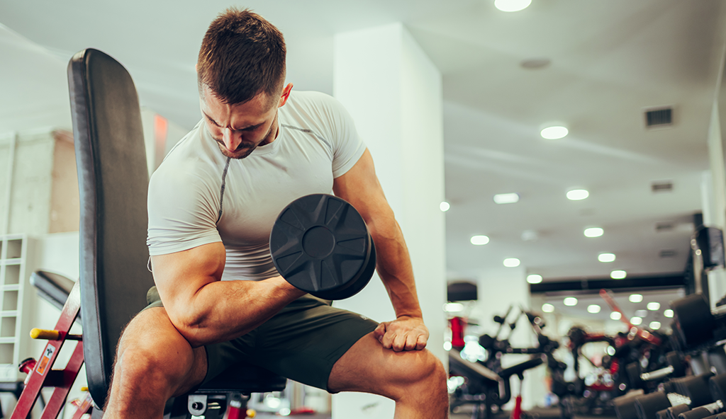
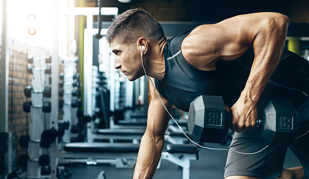
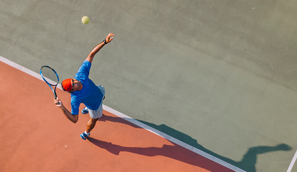
A SLAP Tear (Superior Labrum Anterior Posterior) refers to a specific type of labral tear in the shoulder, which is located at the top of the shoulder socket (glenoid) and involves the attachment site of the biceps tendon. Acute trauma and overuse are often the causes of this specific type of labrum injury.
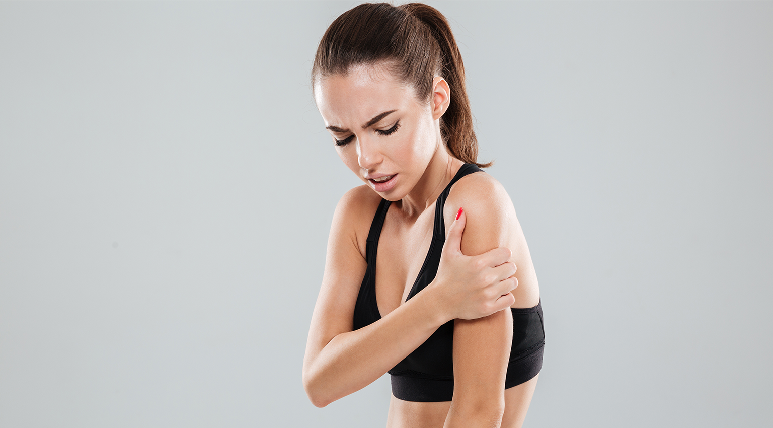
Symptoms

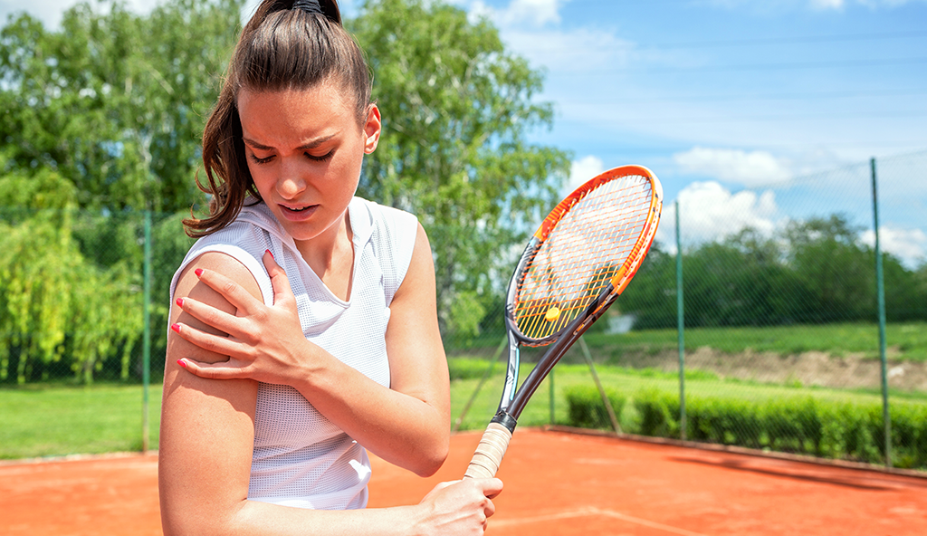
A SLAP tear is often accompanied by pain deep in the shoulder, stiffness, a popping or clicking sensation or feeling of instability. Decreased range of motion or strength can also be present.

Diagnostic Testing

Dr. Anz will conduct a thorough examination and perform a physical examination to help determine whether or not you have a SLAP tear. During the evaluation, Dr. Anz will determine if the tear is associated with any type of pre-existing instability to the shoulder or if it’s from a particular traumatic event. X-rays will rule out any fractures or bone-related issues. Typically, an MRI is most effective in diagnosing a tear.

Treatment

Several key factors play a role in the decision-making process regarding treatment of a tear, including age, type of tear, and athletic profile.

Non-surgical

Initially, patients are directed to rest and ice the area, along with taking anti-inflammatory medications followed by a course of physical therapy.

Surgical

When non-surgical treatments fail, arthroscopic surgery of the shoulder is typically recommended. Generally speaking, there are three surgical options for a SLAP tear

  • Debridement: During a debridement procedure, Dr. Padelecki will smooth out the torn labrum during an arthroscopic surgical approach. This option is only suitable for stable SLAP lesions that do not seem to involve the biceps tendon.
  •  SLAP Repair: A SLAP repair is an arthroscopic procedure that uses anchors with sutures attached to secure the torn labrum down to the shoulder socket. A SLAP repair is the most common procedure done for symptomatic SLAP lesions and is typically reserved for young patients with an otherwise healthy shoulder who want to remain athletically active.
  • Biceps tenodesis: A biceps tenodesis cuts the biceps tendon from where it attaches to the labrum, and reattaches it to another area. By decreasing the forces that pull on the SLAP region, the symptoms will be alleviated. A biceps tenodesis can either be performed arthroscopically, or through a small incision.

Post-Op

A rehabilitation program will be prescribed at your first post-operative visit with Dr. Anz. Rehabilitation after surgery is extremely important, especially in athletic individuals who are seeking to get back into regular activity. Without proper rehab, the chance of full recovery is diminished and shoulder stiffness can occur.

For additional information on labral and slap tears, or to learn more about arthroscopic shoulder surgery, please contact the Gulf Breeze, Florida orthopedic surgeon, Dr. Adam Anz located at the Andrews Institute. 

Shoulder Pain

Frozen Shoulder

Shoulder Pain
Frozen shoulder, also known as adhesive capsulitis, is a condition characterized by inflammation and stiffness in the shoulder joint which can restrict motion and cause chronic pain. Also referred to as “stiff shoulder”, this condition develops when the soft tissue of the shoulder joint begins to thicken and contract as a result of the formation of scar tissue. The scar tissue (referred to as “adhesions”) creates the loss of motion. The condition usually comes on slowly, but generally worsens over time. With the right treatment, frozen shoulder usually can be corrected and the symptoms alleviated.

Symptoms

The most common symptom of frozen shoulder is pain, which is typically described as dull or aching. It is worse early in the course of the disease and is usually located over the outer shoulder area and sometimes the upper arm. Limited mobility, shoulder weakness, and loss of movement of the joint are also major symptoms associated with frozen shoulder.

Diagnostic Testing

Dr. Anz may suspect frozen shoulder during a routine examination and after questioning of your past medical history. An X-ray or an MRI may be performed to see whether symptoms are from another condition such as arthritis, rotator cuff tear, or a broken bone.

Treatment

Non-Surgical Treatment for frozen shoulder usually starts with basic activities including rest, ice, and medication. Physical therapy is often very effective in treating frozen shoulder, especially when combined with steroid injection. These treatments should help the return of motion and function to the shoulder. Surgical If conservative treatments are not curing the frozen shoulder over a period of time, Dr. Anz will perform arthroscopic shoulder surgery to repair the condition. Prior to surgery, he may attempt a manipulation procedure under anesthesia, where he will force the shoulder joint to move, causing the capsule and scar tissue to stretch or tear. This releases the adhesions and increases range of motion. During arthroscopic surgery, Dr. Anz will attempt to cut some of the tight tissues around the shoulder and relieve the pressure.

Post-Op

Following arthroscopic shoulder surgery, patients will need to adhere to a rehabilitation program as instructed by Dr. Anz. This will involve specific exercises and movements over a distinct period of time. It’s in the patient’s best interest to complete rehab and work with Dr. Anz towards a complete recovery. For additional resources on available treatment for frozen shoulder, please contact the Gulf Breeze, Florida orthopedic surgeon, Dr. Adam Anz located at the Andrews Institute. 

Dislocated Shoulder and Shoulder Instability

The shoulder joint offers the largest range of motion in the human body. Made up of three bones: the humerus (arm bone), the scapula (shoulder blade), and the clavicle (collar bone), it is often susceptible to injury because of this great scope of mobility. The shoulder joint is a ball and socket type joint, allowing individuals to perform activities over their head, in front and in back of their body, and using the full rotation that is needed for sports that involve throwing, serving, or overhead function.

The shoulder relies heavily on many soft tissue structures to provide stability: including the muscles surrounding as well as ligaments and the glenoid labrum. With a shoulder dislocation these structures can become injured which may affect subsequent shoulder stability. With a shoulder dislocation, the ball and socket (the humeral head and glenoid) become separated and are not in contact, with the humeral head moving forwards, backwards or downward.

Dislocated shoulders are a common injury among athletes. Individuals who compete in contact and tackling sports such as football, hockey, and rugby are at a higher risk of a dislocation event than non-contact sports. Outside of sports, a traumatic event such as a hard fall can also create this injury. When a young active adult suffers from a dislocation caused by trauma, the risk of a repeat dislocation or future shoulder instability is high.

Symptoms

Symptoms of a shoulder dislocation will vary depending on the severity of the dislocation. Most patients will here a popping sound or sensation, followed by immediate pain. A loss of range of motion will also occur.

Diagnosis

Most individuals who suffer from a shoulder dislocation often have the injury reduced in an Emergency Room, requiring a physician or trainer to maneuver the joint back into place. While this offers a quick fix for the dislocation itself, a thorough physical examination, followed by an X-ray is necessary to evaluate the injury. A MRI should may follow to assess the ligaments and labrum of the shoulder to see if they may have been damaged. Dr. Anz will assess the injury and discuss treatment options and expectations.

Treatment

Treatment for a shoulder dislocation will depend on the severity of the injury. Dr. Anz will take into account the activity level of the patient, including age, number of previous dislocations, and future goals when determining the proper treatment course. If non-surgical treatment is thought to be the best solution, Dr. Anz will recommend ice, rest, immobilization in a sling and physical therapy to help strengthen the nearby structures of the shoulder joint.

Surgical Treatment

Depending on the extent of injury and the number of times of instability, Dr. Anz may recommend surgery. Recurrent dislocations may cause further injury which may increase the chance of further shoulder instability. Arthroscopic surgery can be used to repair the glenoid labrum in episodes of shoulder instability. In some instances, open surgery may be necessary to repair structures and restore stability.

Post-Operative

Following surgery, Dr. Anz will prescribe a thorough rehabilitation program design to restore full function of the shoulder joint. It’s important for patients to understand that for a full recovery, rehab is necessary so that range of motion, strength, and overall mobility can be achieved over time. Most patients are able to resume normal activities within 4-6 months following surgery.

Dr. Anz is an orthopedic shoulder surgeon and sports medicine specialist in the Gulf Breeze/Pensacola, Florida area. If you have sustained a shoulder dislocation, have recurrent shoulder instability, or are seeking additional resources to treat your shoulder pain, please contact our office to schedule a consultation.

Bicep Tendonitis

Biceps Tendonitis

Bicep Tendonitis

Injury Overview: 

Biceps tendonitis of the shoulder is an inflammatory condition of the long head biceps tendon that affects the area where the bicep muscle meets the front of the shoulder. This condition is very common among athletes where extra strain is placed on the biceps tendon. There are multiple reasons why biceps tendonitis may exist. Repetitive motion of the shoulder joint and overuse are usually the main culprits. Multi-directional instability and direct trauma of the joint are other causes.

In some instances, biceps tendonitis can be prevented. Consult with Dr. Anz to review your exercise regimen and discuss strategies to care for your shoulder and prevent overuse injuries.

Symptoms

There are several significant warning signs associated with long head biceps tendonitis. This disease process is most often accompanied by pain in the front of the shoulder associated with movement. This pain will be felt when you extend your arm out in front of you or raise your arm above your shoulder. As biceps tendonitis develops, the pain may become more severe, spread over a larger area and be felt throughout the day. Additionally, patients may feel pain when the actual area is touched and complain of pain radiating down toward the elbow. The area may be red or swollen and you could feel a burning sensation. Some patients feel or hear a snapping sound in the shoulder during movement.

Diagnostic Testing

Dr. Anz will examine your bicep muscle and shoulder for signs of tenderness and inflammation and may then order some tests such as X-rays, or MRI to identify any other problems in the area and confirm the diagnosis.

Treatment

Non-Surgical

Biceps tendonitis is best treated by resting the affected area and slowly working back into light movement. Applying ice to the affected area and taking anti-inflammatory medications may help to alleviate the pain and inflammation. In more severe cases, a corticosteroid injection will help to reduce more acute pain for a longer duration.

Surgical

Surgery for biceps tendonitis is usually performed arthroscopically and is only needed in severe cases where a tendon is damaged. In some cases, the damaged section of the biceps is removed, and the remaining tendon is reattached to the upper arm bone in a procedure called a biceps tenodesis. Removing the painful part of the biceps from the shoulder usually alleviates symptoms and restores normal function.

In severe cases, the long head of the biceps tendon may be so damaged that it is not possible to repair it. Dr. Anz may elect to release the damaged biceps tendon from its attachment with a procedure called a biceps tenotomy.

Post-Op

Depending on the nature of the injury and the exact surgical repair used by Dr. Anz, patients will be prescribed a strict set of rehabilitation guidelines to use with a physical therapist. Physical therapy is mandatory in order to return the patient to optimum function. Most patients have good results and typically regain full range of motion and mobility after completing the program.

For additional information on biceps tendonitis, or to learn more about arthroscopic shoulder surgery, please contact the Gulf Breeze, Florida orthopedic surgeon, Dr. Adam Anz located at the Andrews Institute. 

Bicep Tendon

Biceps Tendon Injuries

Bicep Tendon

Injury Overview

The biceps is located on the front of the upper arm and is made up of two muscle units that originate from the shoulder and insert on a bone in the forearm. The two units of the biceps are known as the short head of the biceps (which attaches to the shoulder blade at the coracoid), and the long head of the biceps (which attaches to the shoulder blade at the top of the shoulder socket). Biceps tendon injury can occur at one of its attachments on the shoulder blade or its attachment in the forearm. The majority of biceps tendon problems occur in the long head of the biceps tendon near its insertion at the top of the shoulder socket, often referred to as proximal biceps tendon injuries.

Injuries to the proximal biceps can range from an inflamed tendon (tendonitis), to a complete rupture. Injuries of this nature are common among individuals who participate in repetitive shoulder activities that involve the use of the arm above the head. In some instances of repetitive stress and irritation, the tendon can become weak and tear partially, causing fraying. In other instances, the tendon can completely tear from its attachment site. A complete tear may occur suddenly during an activity such as heavy lifting. Proximal biceps tendon injuries are also commonly seen in association with tears of the rotator cuff muscles around the shoulder, which may allow the biceps tendon to subluxate (move out of position) from its normal groove.

Injuries to the biceps tendon at the forearm, also known as distal biceps injuries, occur less frequently. Partial or complete detachment of the biceps tendon can occur from the radius, bone of the forearm where the muscle inserts. Often patients report a pop while using their arm for lifting or while their arm was jerked into a straight position. This is often accompanied by swelling, bruising, and a deformity at the arm.

Symptoms

The most obvious symptom associated with a proximal biceps tendon injury, is pain in the front of the shoulder joint with overhead activity. With documented tendon tears, patients often report a sudden, sharp pain and a “pop” feeling or sensation that occurs when the tendon tears. Most patients will report weakness in the shoulder or elbow depending on the location of injury. Bruising, and a change in the appearance of the front of the arm may occur as well. This bulge results as a result of the altered resting position/tension of the biceps muscle and is often called a pop-eye deformity.

Diagnosis

To determine the proper course of treatment, Dr. Anz will perform a physical examination of the shoulder and arm, and will determine the patients level of pain, function, strength, and mobility. Specific physical exam tests for proximal biceps tendon injuries include the Speed’s test, Yergason’s test, and tenderness over the biceps groove. Specific tests for biceps tendon injury at the forearm include the hook test, passive forearm pronation test, and biceps crease interval test. In addition to specific clinical tests, an MRI to confirm a diagnosis may be necessary.

Treatment

With biceps injury at the shoulder, if the diagnosis is consistent with biceps tendonitis or a strained tendon, Dr. Anz will usually recommend non-surgical treatment measures including rest, ice, anti-inflammatory medication, a change in daily activities that irritate the tendon, and physical therapy. In some instances, an injection into the tendon sheath can help decrease the inflamation/irritation at the tendon. In cases of a complete tear of the tendon at the shoulder, many patients still retain good function without surgery. However, some patients may exhibit symptoms of a deformity and cramping pain. In these scenarios, a biceps tenodesis may be appropriate. This involves surgery to reattach the torn tendon at a different location to alleviate cramping pain and restore a normal arm contour.

With biceps injury at the forearm, partial injuries typically do well without surgery. A period of rest and recovery is necessary. Complete tears of the biceps at the forearm are accompanied by a 30% decrease in forearm flexion strength and a 40% decrease in supination strength of the elbow. For some patients, this decrease in strength is acceptable and function/pain will not be a long term problem. For some patients, surgical repair is important due to their occupations and/or recreations.

Post-Operative

In surgical scenarios, Dr. Anz will limit activities during the first portion of the recovery phase. He will also prescribe a thorough physical therapy rehabilitation program that will detail appropriate progress regarding motion and strengthening exercises. It is necessary to allow time for the tendon to heal to bone. It typically takes 2-3 months before patients can return to their normal functional activities.

To learn more about biceps tendon injuries, or for additional resources on arthroscopic shoulder surgery, please contact the Gulf Breeze, Florida orthopedic surgeon, Dr. Adam Anz located at the Andrews Institute. 

Shoulder Arthritis

Shoulder Arthritis

Shoulder Arthritis

Injury Overview

Shoulder arthritis is a relatively common condition in which the joint (ball and socket) becomes painful and dysfunctional as a result of wear and tear in the shoulder. In a normal shoulder, the ends of the bone are covered by a smooth surface known as articular cartilage. In patients with shoulder arthritis, this cartilage is progressively lost, exposing the bone beneath and causing chronic pain and in some cases, loss of movement.

There are 2 distinct joints in the shoulder that can be affected by arthritis—the AC joint (acromioclavicular), where the collarbone (clavicle) meets the bony roof of the shoulder (acromion); and the glenohumeral joint where the ball of the arm bone (humerus) meets the socket (glenoid).

There are several types of shoulder arthritis:

  •  Osteoarthritis is the gradual wearing down of the joint cartilage that occurs predominantly in those of a more mature age, or is the result of overuse in highly active athletes.
  • Post-traumatic arthritis occurs within the shoulder joint after an acute or traumatic injury (such as a blunt force blow to the joint or a hard fall). Even after the injury itself is repaired, the joint can still be susceptible to early arthritis due to mechanical and chemical changes within the joint. This type of arthritis can also develop after a chronic rotator cuff tear.
  • Rheumatoid arthritis occurs when the body’s immune system attacks its own cartilage and destroys it. This is often a hereditary or genetic form of arthritis that tends to affect more women than men.

Symptoms

The predominant symptom of shoulder arthritis is chronic pain, which is derived from the grinding of the bones against one another after the cushioning function of the cartilage is lost. Some patients may experience some weakness or stiffness in the joint as the condition progresses. Night pain and difficulty sleeping are also common symptoms of shoulder arthritis.

Diagnostic Testing

Dr. Anz will conduct a thorough examination of the shoulder joint to find areas of tenderness or pain and to also check overall mobility. A variety of tests may be utilized to confirm the diagnosis of shoulder arthritis, including X-rays (to look at the joint) or an MRI (to assess the soft tissue and cartilage).

Treatment

The goal in treating shoulder arthritis is to reduce or eliminate the underlying symptoms associated with the condition. This includes alleviating the pain, swelling, and overall stiffness of the shoulder joint.

Non-surgical

Arthritis of the shoulder is very common. Many patients are able to live with the side effects for years before finally seeking medical treatment. Most patients find that resting the shoulder and avoiding activities that exacerbate inflammation helps. Applying ice to the joint and taking anti-inflammatory pain medications also help with pain. Corticosteroid injections also are helpful to relieve the symptoms and these may be administered by Dr. Anz. Physical therapy exercises, such as swimming, can be soothing and may help maintain joint motion while strengthening the shoulder and avoiding impact.

Surgical

In patients where the symptoms associated with shoulder arthritis are severe and continue to worsen, there are a number of surgical procedures that exist to help. Treatment for shoulder arthritis is based upon the cause and severity of the arthritis, the intensity of the symptoms, and the functional level of the patient. The effect of this disease on daily life can often be the deciding factor on treatment strategy.

In its early stages, shoulder arthritis can be treated via arthroscopic surgery, which is the least invasive approach. With this operation, Dr. Anz trims out the inflamed synovial lining tissue and removes pieces of the degenerated cartilage. This treatment will not completely cure the arthritis, but can relieve many of the symptoms.

In severe cases, the recommended surgical treatment is shoulder replacement surgery (or joint replacement). This operation often restores motion lost through the degeneration by replacing the damaged ball with a synthetic surface. Arthroplasty is the term generally used for replacement surgery.

Replacement surgery is not recommended for young patients. There are other joint restoration and joint preservation techniques that have been proven effective for younger patients (under the age of 70). Typically, a joint replacement procedure is the final opportunity to treat the arthritis and the longevity of the procedure can last up to 15 years.

Post-Op

A rehabilitation program will be prescribed at your first post-operative visit with Dr. Anz. Rehabilitation after surgery is as important as the surgical repair itself. Without proper rehab, the chance of full recovery is diminished. Depending on the exact surgical procedure that was performed, the patient will be required to do exercises and strengthening moves with a therapist.

For additional resources on arthritis of the shoulder, or to learn more about arthroscopic shoulder surgery, please contact the Gulf Breeze, Florida orthopedic surgeon, Dr. Adam Anz located at the Andrews Institute. 

push-up

AC/CC Joint Injuries

push-up

Shoulder anatomy involves three bones and two important joints:  the scapula (shoulder blade), the humerus (upper arm bone), and the clavicle (collarbone) are the bones of the shoulder.   The two joints of the shoulder are the glenohumeral joint (joint between the humerus and scapula) and the acromioclavicular joint (joint between the clavicle and scapula).  The acromion, from the Greek Akros-shoulder and omos shoulder, is a portion of the scapula which is palpable on the outside portion of the shoulder.  The acromion and clavicle meet above the glenohumeral joint at the acromioclavicular joint, also known as the AC joint for short.  This joint is held together by ligaments between the acromion and clavicle which stabilize the AC joint and are known as AC ligaments.  In addition, important ligaments between the clavicle and coracoid, another portion of the scapula, are also important in stabilizing the AC joint and entire shoulder; these ligaments are known as CC ligaments.  The AC and CC ligaments can become injured during direct contact onto the shoulder, such as when a football player is tackled and lands directly on the side of his shoulder.  Injuries to the AC ligaments and CC ligaments can be associated with displacement of the shoulder blade in relation to the collar bone, an injury sometimes referred to as a shoulder separation.  This injury is not to be confused with a shoulder dislocation, which is when displacement occurs at the glenohumeral joint.

AC joint injuries are graded in severity and labeled Type I-VI.  Type is classified by displacement of the acromion in relation to the clavicle with suspected injury to the AC and CC ligaments based upon this displacement.  Displacement typically involves the acromion below the clavicle; however, on x-ray this is often interpreted as the clavicle being above the acromion.

  • Type I: No Separation on X-ray, pain with palpation at AC joint, mild stretch of AC ligaments
  • Type II: 0-100% separation at AC joint on X-ray, tearing of AC ligaments with stretching of CC ligaments
  • Type III: 100-125% separation at the AC joint on X-ray, tearing of AC and CC ligaments
  • Type IV: Displacement of the clavicle posteriorly on X-ray with injury to the AC and CC ligaments
  • Type V:  Displacement of between 125% to 300% on X-ray with acromion below calvicle.
  • Type VI:  An AC joint separation where the clavicle is displaced 100% or more below the acromium on X-ray. This injury is quite severe and extremely rare

Symptoms

AC joint injuries vary according to the spectrum of damage of the AC and CC ligaments.  Patients who present with Type I and II injuries will experience milder symptoms that include tenderness over the AC joint, minor swelling, pain during movement, and a  mild to moderate deformity that may be visible to the eye. In more severe cases, symptoms will be more extreme and bruising will also appear on the outside of the skin.  Most patients note hearing a popping sound at the time of the initial injury. This is caused by the disruption of the AC joint at the time of injury. A higher-grade injury will almost always result in a prominent bump on the shoulder, severe pain, and limited mobility.

Diagnosis

AC joint injuries are relatively easy to diagnose. However, the severity can vary so greatly that an X-ray or MRI may be necessary to fully determine the extent of injury and treatment recommendation.  During the initial visit, Dr. Anz will examine the shoulder to evaluate the injury. On examination, patients with an AC joint injury usually have tenderness directly over the AC joint and pain when bringing their arm across the body. It is important to determine upon examination if other shoulder structures may have been damaged.  If this is suspected, an MRI is important to evaluate for associated injury.

Treatment

Dr. Anz will attempt to treat the injury in the most conservative way possible. For minor AC joint injuries where a mild sprain or low-grade separation exists, he will prescribe anti-inflammatory medications, a sling for comfort, and a protocol of rest with therapy for rehabilitation when appropriate. A majority of the milder injuries can successfully be treated non-operatively (without surgery).

Surgical Treatment

For higher grade AC joint injuries, surgery may be needed in order to repair or reconstruct the ligaments involved in the injury and address nearby structures. The ultimate goal during surgery to treat the AC Joint is to stabilize the joint and correct the separation of the clavicle and acromium.  Dr. Anz will discuss surgical options with patients and agree upon the most appropriate technique when necessary.  Open stabilization and arthroscopically assisted stabilization techniques are both used depending on the clinical scenario.

Post-Operative

Following shoulder surgery of any kind, Dr. Anz will prescribe a thorough and detailed post-op rehabilitation program.  Rehab following an AC joint surgery will often span about three months during which physical therapy incorporating strengthening exercises will be slowly incorporated. Physical therapy is a team effort between Dr. Anz and his patients so that an optimal outcome can be achieved. Most athletes are able to resume their sports within 4-6 months following surgery.

For additional information on AC joint injuries and shoulder separations, or to learn more about arthroscopic shoulder surgery, please contract the office of Dr. Adam Anz, orthopedic shoulder surgeon in Gulf Breeze, FL.

External Snapping Hip Syndrome

Trochanteric Bursitis and External Snapping Hip Syndrome

External Snapping Hip Syndrome

Injury Overview

Bursal tissue is present in the body to decrease friction between two adjacent moving structures. When this tissue becomes irritated (or inflammed), the condition is known as bursitis. The greater trochanter bursa lies between a tendon-like structure in the thigh known as the iliotibial band and a boney prominence in the thigh known as the greater trochanter. Greater trochanteric bursitis (sometimes referred to as trochanteric bursitis) can be caused by different things in all population types and may cause significant pain. Sometimes greater trochanteric bursitis is caused by changes in a person’s walking, running, or exercise mechanics. As we get older these mechanics change as our muscle bulk and posture change. In addition to changes in posture and mechanics, bursal tissue may also become inflammed in scenarios of overuse or mechanical irritation such as in the setting of external snapping hip syndrome. External snapping hip syndrome occurs when the iliotibial band continually snaps over the greater trochanter. This can occur if the iliotibial band is too tight or too thick.

Symptoms

Symptoms of trochanteric bursitis of the hip may vary from patient to patient. Often the main symptom is pain located on the outside portion of the thigh near the boney prominence of the greater trochanter. Often patients have trouble sleeping on their side and have reproducible discomfort with pushing on the greater trochanter. Pain may also occur when an individuals attempt to run or rotate their thigh/leg. Pain can also be present when one attempts to stand after a long period of sitting.

External snapping hip syndrome is marked by a popping sensation as a patient bends and straightens their hip. As patients flex their hip, the iliotibial band slides toward the front of the thigh over the greater trochanter. As patients extend their hip, the iliotibial band slides toward the back of the thigh over the greater trochanter. In patients with external snapping hip syndrome, this sliding is accompanied by an audible or palpable “pop” which can be painful. Sometimes, patients feel that this is a sign that their hip is popping into and out of joint. However, during this process the ball and socket of the hip joint remains as it should in place.

Diagnosis

Dr. Anz may first order X-rays to rule out other bony abnormalities that may be the source of pain or snapping. He will then obtain a history and perform an examination, including the patients’ discription of the pain/symptoms and a history of sports related injuries and activity. He will examine the lower extremities in order to see which movements cause discomfort and will also perform certain physical examination tests. In some scenarios, a MRI may be important in order to rule out other injuries or ensure a correct diagnosis. Relief of pain with an injection of an anesthetic into the bursa may also be used to confirm that bursitis is the primary source of pain.

Treatment

In most circumstances trochanteric bursitis does not require surgery. Injections in addition to physical therapy is often effective, especially when bursitis is caused by hip girdle weakness or changes in posture. There are four major stabilizers of the hip joint including the muscles of the hip girdle. Therapy focusing on pelvic girdle strengthening, core strengthening, and stretching of the ITB band can often make a significant improvement in mechanics and take pressure off of the bursa. Additionally, anti-inflammatory medications (taken by mouth or rubbed onto the region) and rest from aggravating activities can greatly benefit the patient and relieve associated symptoms.

Surgical

In rare cases, Dr. Anz may recommend surgical treatment. Surgery for the treatment of bursitis is most effective when a mechanical irritating force can be removed or an inciting mechanical scenario improved, such as in the setting of external snapping hip syndrome or femoroacetabular impingment. Dr. Anz will use an arthroscopic approach in order to remove the inflamed bursa and loosen the iliotibial band. During arthroscopic surgery, he will make keyhole incisions through which he will enter small surgical instruments and a camera. He will remove the inflamed bursa and lengthen the IT band in order to relieve pressure.

Post-Operative

Following arthroscopic surgery for trochanteric bursitis, Dr. Anz will prescribe a brief period of rest followed by a physical therapy program to help restore strength and motion. Patients often make great progress during physical therapy and can resume normal activities within 6 to 8 weeks.

For more information on trochanteric bursitis of the hip, or if you have any other questions regarding surgical treatments for the hip, please contact the Gulf Breeze, Florida orthopedic surgeon, Dr. Adam Anz located at the Andrews Institute. 

PVNS Pigmented Billonodular Synovitis

PVNS

PVNS Pigmented Billonodular Synovitis

Injury Overview

Pigmented villonodular synovitis, or PVNS, is a disease caused by the abnormal growth of the hip joint’s lining tissue, known as the synovium. Excessive growth of this tissue leads to inflammation in the joint causing pain, tenderness, and stiffness. The condition occurs most commonly in patients in their 30s and 40s, but the actual cause of PVNS is still unknown.

PVNS is identified in two different forms:

  • Localized – Pain and swelling occurs in just one area of the joint, and responds well to treatment.
  • Diffuse – More common than localized PVNS, diffuse PVNS involves the entire joint, and can be more destructive and difficult to treat.

Symptoms

Symptoms of PVNS include hip pain, weakness, and stiffness of the joint. Patients with localized PVNS may experience joint locking, a catching sensation or a feeling of instability. Patients with diffuse PVNS may experience a gradual onset in symptoms.

Diagnostic Testing

PVNS may not necessarily be evident on a standard X-ray, though an X-ray could show other potential problems that could be causing the hip pain. Dr. Anz may choose to order an MRI scan that will more accurately show evidence of a nodular mass with bone changes if the condition is localized PVNS. If the condition is diffuse PVNS, an MRI will show a thickening of the joint lining or an extensive mass, potentially with destructive bone changes.

In some cases Dr. Anz may perform a procedure called a joint aspiration, whereby he releases fluid from the hip joint. Typically with PNVS cases, the joint fluid appears bloody. A biopsy is the confirmatory test for PVNS of the hip.

Treatment

Surgery is the best option to treat a diagnosis of PVNS. Dr. Anz will remove the painful, inflamed synovial lining through a procedure called a synovectomy, which, can be done via arthroscopic or open surgery.

In an arthroscopic synovectomy, Dr. Anz surgically removes the affected lining with the mass only using small incisions. This is the most common way to treat localized PVNS. However in the instance of diffuse PVNS, a total synovectomy may be required, which is an open surgery procedure that removes the mass and the entire joint lining.

Post-Op

Following a synovectomy, patients should avoid full weight bearing as discussed with Dr. Anz at the first post-operative visit. For patients recovering from arthroscopic procedures, the recovery time is shorter, and will involve a short course of physical therapy before resuming normal daily activities. However for those who undergo the open procedure, a more extensive physical therapy program will be prescribed to prevent post-op stiffness and a return to normal mobility, often taking months.

For more information on PVNS (pigmented villonodular synovitis) or other disorders of the hip, please contact the Gulf Breeze, Florida orthopedic surgeon, Dr. Adam Anz located at the Andrews Institute. 

Internal Snapping Hip Syndrome

Internal Snapping Hip Syndrome/Psoas Impingement

Internal Snapping Hip Syndrome

Injury Overview

A condition of the hip which can affect recreational and professional athletes alike is internal snapping hip syndrome. This syndrome is marked by snapping of the iliopsoas over the front of the hip joint. The iliopsoas is a muscle, also referred to as the hip flexor, which connects the bone in the thigh (femur) to the lumbar spine. It starts as a muscle at the lumbar spine and transitions to a tendon as it courses to its attachment on the femur. At the hip joint, there is a significant portion which is tendon and a significant portion which is still muscle. The tendon portion often runs directly over the front of the hip joint, and sometimes this tendon can create a popping sensation as it moves across the hip joint. Some patients describe this popping as their hip popping out of joint, but most often this popping represents motion of the tendon alone. In some instances, the muscle and tendon of the hip flexor can become tight and irritated (inflammed). This is often associated with groin pain and a snapping sensation which can be quite bothersome. In severe instances, a tight iliopsoas with long standing popping can lead to inflammation and/or damage of the acetabular labrum, also creating hip pain and irritation. This is condition is known as psoas impingement. Without proper treatment and rest, the labrum can become damaged to the point where it tears.

Symptoms

Internal snapping hip syndrome is a precursor to psoas impingment. Symptoms of internal snapping hip syndrome include popping located in the groin. At times the popping can be accompanied by a sharp pain. With time as the snapping progresses to psoas impingement a constant, deep ache within the groin may develop. Patients may also feel tightness along the front portion of the hip joint. Often symptoms associated with internal snapping hip syndrome and psoas impingement are increased with activities and sports.

Diagnosis

Dr. Anz obtains X-ray’s of the hip and pelvis to evaluate a given patient’s anatomy and look for abnormalities which can predispose one to hip impingement. He will obtain a comprehensive history to consider what types of activities you participate in and how they may affect your hip. He will conduct a physical examination that will evaluate the status of your hip flexor. The Thomas test is a physical examination test that will allow him to determine how tight a patient’s iliopsoas tendon/muscle is. He will also listen for any sound that the joint may make during his examination. If appropriate, he will order an MRI to assess the iliopsoas as well as the acetabular labrum.

Treatment

Upon diagnosis of psoas impingement, Dr. Anz will begin to discuss treatment options. In most situations, a patient can recover fully from impingement without the need to have surgery. Non-surgical treatment techniques which Dr. Anz will recommend include rest, the use of anti-inflammatory medications, physical therapy, and/or injection of steroids into the tendon sheath to decrease inflammation and alleviate pain. A thorough stretching and strengthening program is often the best first step to making patient’s better. A therapist is often necessary to help assist with this progress. If these treatment techniques are attempted and are not successful, Dr. Anz may begin to discuss surgery with the patient.

Surgical Treatment

If surgery is required to treat psoas impingement, Dr. Anz will perform it arthroscopically. During an arthroscopic surgery Dr. Anz will insert a camera and small surgical instruments through 2 to 3 small incisions along the hip. If FAI impingement is also involved, he will address this and any damage created at the same time. Releaseing the psoas tendon, medically known as “fractional lengthening,” involves cutting the tendon portion at the hip joint while preserving the portion which is still muscle at the hip joint. Fractional lengthening may cause some future weakness in hip flexion strength.

Post-Operative

Physical therapy is a crucial part of the healing process for patients in order to make a full recovery. A progressive rehabilitation program will be prescribed at your first post-operative visit with Dr. Anz. Initially, the therapy will focus on slowly returning motion back to the injured hip. After motion is regained, patients will follow a progressive strengthening program, and eventually, a return to normal activities.

If you believe you are suffering from Psoas impingement, or would like additional information on the causes of hip pain, please contact the Gulf Breeze, Florida orthopedic surgeon, Dr. Adam Anz located at the Andrews Institute.