Multi Ligament Knee Injury

Multi-Ligament Knee Reconstruction

Multi Ligament Knee Injury

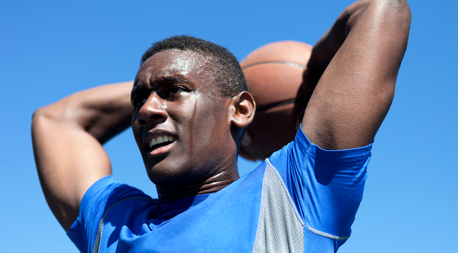
Multi-ligament knee injuries are serious injuries that are often considered complex.  This type of injury occurs when more than one ligament is affected during injury.  There are four major ligaments that make up the knee joint. Each has their own function but together they work to stabilize the knee.   

These ligaments include:

  • The anterior cruciate ligament (ACL)
  • The posterior cruciate ligament (PCL)
  • The Medial collateral ligament (MCL)
  • The Posterior lateral complex (PLC)

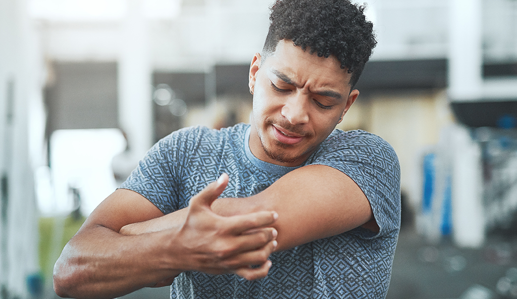
Multi-ligament knee injuries can occur as the result of a traumatic incident such as a collision or fall during sports, an automobile accident, or a serious fall such as snow skiing. Each injury is different and depending on different mechanisms, a multitude of injury patterns can be present.

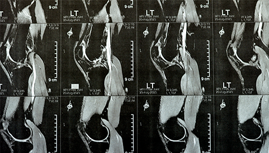
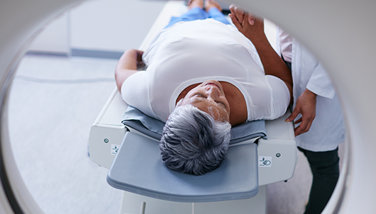
In many instances where multiple ligaments are affected, a multi-ligament knee reconstruction would most likely need to take place.  An MRI will be able to detail the extent of the ligament injury. When multiple ligaments are involved, there are occasions where one ligament can heal on its own while another is reconstructed, and there are occasions where more than one, or multiple, ligaments must be reconstructed.

Multi-Ligament Knee Injury

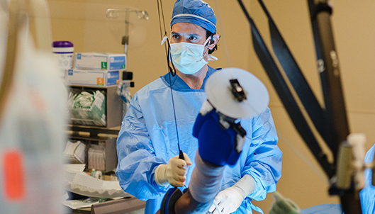
Reconstruction of ligaments such as the ACL and PCL requires removing the damaged ligament and reconstructing a new ligament with a graft.  This graft may be from the patient’s own tissue (known as an autograft), or it could be from donated tissue from another person (known as an allograft).  In some instances, it may be necessary to use multiple allografts. The goal of this surgery is to attempt to secure the ligaments back to their native position.  Dr. Anz will often perform this surgery in one operation and once the ligaments are attached back to their anatomic sites, during early post-op, one can start early range of motion to minimize the chance of the patient developing stiffness and scaring around the knee.

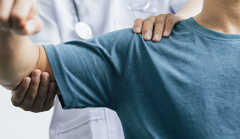
Following this surgical procedure, physical therapy will begin immediately; however, knee range of motion may be limited for a period of time.  Bracing and progression with therapy is individualized based upon the type of injury. Multi-ligament knee surgeries are serious procedures and a thorough, and intensive rehabilitation program will need to be followed post-op in order to obtain the most optimal results.

For additional information on multi-ligament knee injuries, or to learn more about multi-ligament knee reconstruction, please contact the Gulf Breeze, Florida orthopedic surgeon, Dr. Adam Anz located at the Andrews Institute.

A golfer takes aim on the links.

MCL Reconstruction

A golfer takes aim on the links.

The MCL (medial collateral ligament) is one of the four major restraints to the knee joint. 

Along with the anterior cruciate ligament (ACL), posterior cruciate ligament (PCL), and posterior lateral complex (PLC), it works to provide stability and strength to the knee joint.  The MCL is the main ligament on the inside of the knee, also known as the medial side, and is a part of the “posteromedial corner” (PMC).  This ligament can become injured with certain twisting injuries. Often, the MCL will heal on its own after injury; however, in certain instances the ligament is torn to the extent that an MCL reconstruction is necessary.

By examining a knee and obtaining X-rays where the ligament is stressed, it is possible to determine which injuries will heal without surgery and which will require surgery. Whether the MCL tear is partial or complete, Dr. Anz will be most concerned about the overall stability of the joint. During his initial evaluation, he will also determine if the MCL was injured along with the ACL, which is commonly the case. MCL reconstruction involves harvest of one of the hamstring tendons.  This tendon is used to reconstruct the damaged MCL.

After surgery, patients will be required to undergo rehabilitation involving physical therapy in order to obtain an optimal result. Physical therapy begins immediately; however, knee range of motion is limited for 2 weeks.  

MRI of left knee joint showing minimal joint effusion, PHMM Posterior Horn Medial Meniscus degeneration, ACL anterior cruciate ligament mild sprain, normal MCL, LM, LCL, ligaments, vessels and nerves

Initially, patients cannot place weight on their leg for 6 weeks, this amount of time is necessary for ligament healing.  Strengthening begins at 7 weeks, and patients typically return to jogging at 4 months and sports at 5-6 months, depending on the severity of the initial injury.

For additional information on MCL knee injuries, or to learn more about MCL reconstruction surgery using arthroscopy, please contact the Gulf Breeze, Florida orthopedic surgeon, Dr. Adam Anz located at the Andrews Institute. 

Articular Cartilage Defect Surgery

Articular cartilage is a very important component of a healthy knee joint. It covers the ends of each of the bones and is what allows for a fluid, smooth, painless motion of the knee. When an area of the cartilage becomes damaged, a patient will experience swelling and pain with activities, and over time, this process will continue to progress which eventually leads to complete loss of cartilage down to the bone.  When this happens, a diseases known as osteoarthritis becomes present. This continues to cause progressive symptoms of knee pain and stiffness.

When a patient presents with damage to the articular cartilage, or an articular cartilage defect, it is of the utmost importance to treat this appropriately in an effort to restore the damaged area and preserve the surrounding cartilage. This treatment will depend on many factors including size of the defect, depth, location, associated injuries in the knee, age, and activity goals. In many cases with a focal cartilage defect, surgery is recommended.

There are a variety of surgical procedures that can be used to treat articular cartilage damage. These include chondroplasty, Microfracture, osteochaondral autograft transfer, or osteonchondral allograft transplantation.  Listed below are some of the specific behind each technique:

  • Chrondoplasty is an arthroscopic procedure used on some patients which involves using a camera and small instruments to trim away the damaged area of cartilage to alleviate the patient’s symptoms and prevent further propagation of the defect. This procedure is typically undertaken in patients who do not have full thickness defects (down to bone).
  • Microfracture is a technique used in patients who have a full thickens loss of cartilage.  It is a technique where a small pick is used to make holes in the bony surface of the defect. This allows the flow of marrow elements and stem cells which form a clot over the defect and eventually cover the area with a fibrocartilage layer.
  • Osteochondral autograft transfer and osteonchondral allograft transplantations are two types of procedures where an autograft (taken from the patient) or allograft (taken from a donor) is used. In autograft OATs, small plugs of cartilage and bone are taken from another less important area of the patient’s knee and transferred in to fill the defect. In allograft OATs, the cartilage and bone are taken from a donor and also used to fill the defect. These procedures are done with a combination of arthroscopic and open techniques.

Physical therapy will be prescribed following surgery for an articular cartilage defect. Post-operative management varies depending on the specific type of procedure and the severity of the injury. For chondroplasty, patients are typically on crutches for a few days and allowed to weight bear as tolerated with full range of motion. For the other procedures the patient is placed into a brace and weight bearing is protected for 6-8 weeks. Motion may be initially restricted and a continuous passive motion machine (CPM) is also typically used for 6 weeks after surgery.

For additional information on osteoarthritis of the knee, or to schedule an appointment to discuss articular cartilage defect surgery or other treatments for this specific condition, please contact the Gulf Breeze, Florida orthopedic surgeon, Dr. Adam Anz located at the Andrews Institute. 


Meniscus repair and Partial Meniscectomy

Meniscus is a very important part of the knee joint. It is a type of cartilage that serves as a shock absorber within the knee since very high loads are transmitted across the knee with walking, running, jumping, going up and down stairs, or participating in sports or other active extracurricular activities. The menisci are c-shaped pieces of cartilage that cover the knee and are extremely important to distribute the load across the knee and protect the articular cartilage. The menisci also serve as secondary stabilizers to the knee to assist the ligaments.

The knee needs the meniscus in order to function. If a meniscus sustains a significant tear, it loses its ability to function. Over time this will lead to degradation and wear of the articular cartilage, called arthritis.  This eventually can lead to osteoarthritis and bring a myriad of unwanted symptoms.

The symptoms of a meniscus tear include pain on the inside or outside of the knee, a feeling of instability, locking, catching, and tenderness. An MRI will be used to confirm a meniscus injury. If the meniscus shows a small tear, the patient may be able to heal without surgery by way of physical therapy. For larger tears, those that have failed non-operative management, and those with frank mechanical symptoms surgery is typically recommended.

A meniscectomy will be performed on the knee if the meniscus has a small symptomatic tear, one that is shredded beyond repair, or a tear in the inner third of the meniscus. This procedure essentially removes the area of the tear, and removes the damaged section. This is called a partial meniscectomy. Dr. Anz will take great care to only remove the damaged part of the tear and leave as much of the healthy meniscus as possible.

For tears that occur in the outer two thirds of the meniscus, or for larger tears that can be fixed, an attempt is made to repair the meniscus and preserve as much of the native meniscus as possible. This is performed with a combination arthroscopic and open procedure. Strong stitches are placed through the tear to bring the tissue back together and complete the repair.

After a meniscal repair, the knee must be protected to allow the meniscus to heal. For a partial meniscectomy procedure, the patient is allowed to weight-bear as tolerated. Physical therapy is started immediately to regain motion and strength. Weight bearing is protected on crutches for two weeks followed by full weight bearing in the brace, which is worn for a total of six weeks.

To learn more about mensical injuries of the knee, or for additional information on meniscus repairs or meniscus surgery of the knee, please contact the Gulf Breeze, Florida orthopedic surgeon, Dr. Adam Anz located at the Andrews Institute. 


Shoulder Replacement

Shoulder Replacement Surgery

Shoulder Replacement

Osteoarthritis of the shoulder can be a debilitating condition among the older adult population.  

Typically beginning as arthritis, it can take years of wear and tear to cause osteoarthritis and the symptoms that can at times almost be too much to take for some patients. For these patients, once conservative and joint preserving options have been exhausted, shoulder replacement surgery is often recommended.

Shoulder replacement surgery, also known as shoulder arthroplasty, has come a long way in recent years and many patients are able to sustain normal lives following the procedure. During replacement surgery, the natural degenerated cartilage and bone of the shoulder joint are replaced with metal and plastic components.  The metal and plastic components are kept in place with a combination of medical bone cement and tightly fitting the components in place.  These components allow a painless motion of the shoulder after surgery.

It’s important to note that replacement surgery, while it often brings similar, positive results for patients, may not have forever lasting effects.  Most patients are able to live pain-free and active for years following this surgery, however, it is a procedure that is not typically recommended for patients under the age of 65 because it often is considered a last resort procedure to treat osteoarthritis.

A patient enters the MRI portal.

Shoulder replacement surgery will require physical therapy and rehabilitation so that the shoulder joint can regain strength, motion, and overall mobility. Initial therapy focuses on safe motion with certain restrictions for 6 weeks.  

After a recovery period of 2-3 months, patients typically are painless and performing well with their normal activities.

For more information on the treatment of osteoarthritis of the shoulder, or to learn more about shoulder replacement surgery, please contact the Gulf Breeze, Florida orthopedic surgeon, Dr. Adam Anz located at the Andrews Institute. 

Anthroscopic Revision Rotator Cuff Repair

Arthroscopic Revision Rotator Cuff Repair

Anthroscopic Revision Rotator Cuff Repair

The rotator cuff muscles are a very important part of the shoulder joint because they are responsible for all forms of rotation and provide the shoulder the strength it needs in order to perform certain activities.

The rotator cuff is a series of four muscles that make up, and surround the shoulder joint – each one responsible for centering the ball in the socket of the shoulder and providing strength to move the arm and participate in overhead activities.

The muscles that make up the rotator cuff are surrounded and connected by tendons.  Individuals who partake in constant shoulder rotations (such as swimmers), who are injured in sports, or those who experience a traumatic injury such as from a fall or accident, risk damaging these tendons.  This damage may be in the form of inflammation from overuse or bursitis, or from an impingement.   Degenerative wear and tear from chronic overuse can also be the cause for injury. There are a variety of conditions that can occur from damaged rotator cuff muscles-these conditions usually involve a tear in the muscle or tendon.  Depending on the exact injury, treatment can help alleviate the pain and restore function to the shoulder joint.

In the case of rotator cuff injuries where a partial tear, or complete tear, of the muscles or tendons exist, surgery is usually needed to correct this injury. Dr. Anz typically uses an arthroscopic approach to repair rotator cuff tears. In some circumstances a patient may have a re-tear of the rotator cuff after a previous repair. This is often do to some sort of traumatic occurrence which leads to re-injury (and in many cases, is a sign that the patient may have returned to activities too quickly after the initial surgery.)

In these cases, a revision rotator cuff repair is typically required to provide the patient with an optimal outcome. A revision repair is a more difficult procedure due to the complexity of dealing with a tendon that has now torn multiple times and the fact that a repair has already been completed previously.

Dr. Anz conducts revision rotator cuff repairs via an arthroscopic approach as well. During surgery, his primary focus is to use as much of the healthy tendon as he can and reattach it to its native footprint. Some cases may involve a graft or additional attachment to assist in this process. Dr. Anz may also use assisted techniques such as a marrow stimulation healing response or platelet rich plasma.

The revision surgery, and the intricate nature of how it is performed will determine on the exact type of tear or re-tear. In very severe cases involving large, traumatic and chronic re-tears, full repair may not be achievable. These cases are treated with partial repairs, augmentation, or debridement.

After revision rotator cuff surgery, Dr. Anz will prescribe a very detailed, thorough physical therapy and rehabilitation program. The patient’s arm will placed in a sling and therapy will start almost immediately after surgery. The specific progression of rehabilitation will depend on the injury, type of repair, nature of the surgery, age, and overall health of the patient.  Initially, the patient will need to remain in the sling for a period of 6-8 weeks. Therapy typically involves passive range of motion moves, followed by active motion, strengthening, and a slow, steady return to activities.

For additional information on rotator cuff repairs or revision rotator cuff repairs, or for additional resources on rotator cuff injuries and other shoulder conditions, please contact the office of Dr. Adam Anz, orthopedic shoulder surgeon in Gulf Breeze, Florida at the Andrews Institute.


Bicep Pain

Biceps Tenodesis

Bicep Pain

The biceps is a muscle (also referred to as the brachii muscle) that is located on the upper arm between the shoulder and the elbow.  It is the muscle that is responsible for elbow flexion as well as rotating the forearm.

Two muscle “bellies”, make up the biceps region, these include the long head of the biceps and the short head of the biceps (brachii is a Latin phrase which means two-headed muscle of the arm). The long head of the biceps attaches to the shoulder blade inside the shoulder joint through a tendon.  One common injury that can occur to this area is tendonitis.  Biceps tendonitis occurs when acute episodes of overuse takes places. It can also develop when chronic micro-damage from repetitive overhead activities take place, or through degenerative changes in the shoulder joint. Tendonitis occurs when the long head of the biceps muscle becomes irritated.  Symptoms associated with this condition include pain and tenderness in the front of the upper arm.

Dr. Anz will often try and treat biceps tendonitis through conservative measures first, which usually involves rest, ice, anti-inflammatory medications, and therapy. However, if biceps tendonitis causes extreme pain and disability without response to initial treatment, surgical intervention may be the next best step.

If surgery is required, Dr. Anz will most likely perform a biceps tenodesis which is performed arthroscopically.  During this operation, a release of the tendon from its attachment inside the shoulder joint takes place, and it is reattached to the upper arm. The actual release of the tendon is known as a tenotomy.  During the arthroscopic approach, Dr. Anz will make small incisions and use tiny instruments including an arthroscopic camera. The tenodesis is performed through a small incision near the front part of the armpit. Patients can have chronic pain in the front of their arm from biceps tendonitis.  After this tendon is reattached away from the area of previous pain through tenodesis, most patients will feel great relief.

Dr. Anz will prescribe a full rehabilitation program for each patient following surgery. Each case may be slightly different, but the ultimate goal is to regain strength and range of motion. Depending on the severity of the injury and surgical case, range of motion can usually begin shortly after surgery and a full recovery can be anticipated at approximately 3 months.

For additional resources on injuries associated with the biceps muscle, including biceps tendonitis, or to learn more about the surgical treatments for this injury including a biceps tenodesis, please contact the Gulf Breeze, Florida orthopedic surgeon, Dr. Adam Anz located at the Andrews Institute. 


Shoulder Pain

Subacromial Decompression

Shoulder Pain

Patients that experience shoulder pain and shoulder weakness due to bursitis are candidates for a treatment known as subacromial decompression. 

Bursitis and impingement can lead to pain, disability, and tears of the rotator cuff.  The rotator cuff is a group of four muscles that are integral for shoulder function.  These muscles arise from the shoulder blade and attach to the humerus.  As the muscles travel to attach onto the humerus, they travel underneath a portion of the scapula known as the acromium. In some instances, the bursal tissue located in this region can become irritated and inflamed, also known as bursitis.  Additionally, as changes occur with age and use of the shoulder, the acromium may impingement on the rotator cuff.

If conservative treatments fail to work, surgical intervention using an arthroscopic approach can be applied. The surgical procedure to remove the inflamed bursa from the acromium is known as a subacromial decompression.  Using arthroscopic instruments, Dr. Anz will make a small incision in the shoulder and the inflamed portion of the bursa and degenerative portions of the acromium, which are causing the impingement, are removed. Depending on the injury, if bone spurs exist, they will also be removed.

Following subacromial decompression shoulder surgery, patients will be prescribed a physical therapy program. Initially after surgery, they will wear a sling, but will begin active movement of the shoulder pretty quickly. If only a subacromial decompression is performed, range of motion can begin immediately after surgery and a full recovery can be anticipated at approximately 2 months.  Most patients are able to return to their previous activities without pain.

For additional information on shoulder bursitis, impingement, and other conditions that cause shoulder pain, or for additional resources on subacromial decompression, please contact the Gulf Breeze, Florida orthopedic surgeon, Dr. Adam Anz located at the Andrews Institute. 

Shoulder Exercise

Arthroscopic Stabilization for Shoulder Instability

Shoulder Exercise

Shoulder dislocations are the result of the humeral head (ball) and glenoid area of the scapula (socket) to become pulled apart.  Athletes who perform powerful overhead motions, such as serving in tennis or pitching in baseball, put the shoulder joint at risk for dislocations. While the shoulder joint offers the greatest range of motion of any joint in the human body, it also offers an extreme mobility that comes at the expense of stability.

To allow for such great motion, the shoulder is stabilized by soft tissue restraints (such as ligaments and cartilage that surrounds the socket called the labrum). When a shoulder dislocation occurs, the ball comes out of the socket and in most cases, the soft tissue stabilizers are damaged as well.

While the initial treatment for a shoulder dislocation is to reduce the joint (put the ball back into the socket), ongoing instability most likely will occur if further assessment is not performed on the joint to ensure that additional damage doesn’t exist.  Once the shoulder is reduced, X-rays will then be utilized to assess any nearby damage to other bones. Dr. Anz will also determine if the patient should be treated surgically or non-surgically in order to stabilize the joint. If he feels a surgery needs to be performed, he will perform arthroscopic stabilization surgery for shoulder instability.

In the majority of patients, arthroscopic stabilization has been shown to be highly effective in eliminating shoulder instability. In certain situations such as longstanding instability, bone loss from the glenoid or humerus, and a dislocation that can’t be manually reduced, a specialized open procedure may be necessary.

During arthroscopic stabilization surgery, the shoulder is examined to confirm the direction and degree of instability. Next, the area of damage will be assessed and small surgical instruments used to place anchors into the bone on the glenoid that contain strong sutures. These sutures are then used to repair the torn labrum and ligaments, restore the anatomy of the joint to its natural position, and to effectively “tighten” the shoulder back to normal.

Dr. Anz will require all patients to become involved in a post-operative therapy program following arthroscopic stabilization shoulder surgery. This typically consists of gentle passive range of motion movements, followed by active motion, strengthening, and eventually, the return to activities. The patient will continue to wear a cling for about 6 weeks.  Dr. Anz will examine the joint to assess how the progression of therapy should continue.  This will depend on the configuration of the injury and type of repair.

For more information on arthroscopic stabilization shoulder surgery, or to learn more about your specific shoulder injury and shoulder pain, please contact the Gulf Breeze, Florida office of orthopedic shoulder surgeon, Dr. Adam Anz located at the Andrews Institute. 


Shoulder Athlete

Arthroscopic AC Repair

Shoulder Athlete

The collarbone (clavicle) attaches to the roof of the shoulder (acromion) in a joint referred to as the acromioclavicular joint. The collarbone is also stabilized in this area by ligaments called the coracoclavicular ligaments, which attach the collarbone to the front of the shoulder blade (scapula). Direct trauma to this area (such as a football hit or falling over handlebars on a bike) can disrupt these connections and lead to a scenario where the collarbone and roof of the shoulder are no longer sitting next to each other. This is often referred to as an AC separation and leads to elevation of the collarbone, that can often be felt at the top of the shoulder.

Mild AC separations can often be treated by rest and sling use, followed by a short physical therapy program. However, in cases of severe separation, surgery is often warranted because chronic shoulder instability and frequent separations and dislocations can occur on a daily basis following the initial injury. Thus, the goal of surgery is to secure the collarbone back into its normal position by attaching very strong sutures to the collarbone and front of the shoulder blade. This is often accompanied by reconstruction of the coracoclavicular ligaments, which involves looping a donated graft from the front of the shoulder blade to the top of the collarbone. Dr. Anz performs this surgery with the use of the arthroscopic camera as well as a small incision at the top of the shoulder.

Following surgery, patients will start shoulder motion under the direction of a therapist. Patients will be asked to wear a sling for protection for many weeks following surgery. Eventually, after the ligaments heal, patients will be allowed to progressively strengthen the shoulder and discard the sling. Return to sporting activities usually occurs around 3 to 4 months after surgery.

For more information on AC joint injuries, or to learn more about arthroscopic AC repair, please contact the office of Dr. Adam Anz,  Gulf Breeze, Florida orthopedic shoulder surgeon.