Andrews Institute Exterior

Surgery Information

Andrews Institute Exterior

The Andrews Institute is a state-of-the-art facility that utilizes advanced medical tools and equipment for each surgical procedure. 

If during your consultation with Dr. Anz it has been determined that a surgical treatment will be your next step towards treating your condition or injury. W lole offer our patients an organized and relaxed setting so that your surgery takes place on time and without worry. Our patients are always cared for by board certified anesthesia doctors and trained nurses who specialize in regional anesthesia to minimize postoperative pain.

Dr. Anz will discuss the surgical technique with his patients in detail and will also provide a specific set of guidelines for pre-operative, post-operative and rehabilitation. Before surgery, you will receive a physical examination that may include blood work, EKG’s and X-rays. If these routine tests are required, they will be performed a few days before your scheduled surgical procedure.

Below, you will find the most recent pre-operative and post-operative forms for Dr. Anz:

Post-Operative Forms:


Orthopedic Surgery Dr. Adam Anz

For additional information, or if you would like to schedule an appointment please contact the Gulf Breeze, Florida orthopedic surgeon, Dr. Adam Anz located at the Andrews Institute. 

Medical Insurance Dr. Adam Anz

Payment and Insurance

Medical Insurance Dr. Adam Anz

Dr. Anz requests that payment be made at the time of service.  Our office accepts many insurance policies and we will collect the necessary co-pay also at the time of service.  Since the majority of insurance plans do not reimburse the full amount due, you will be responsible for any remaining balance not covered under your plan.   If you have any questions regarding your coverage or account balance, please contact our business office at 850-916-8463.

Dr. Anz is proud to accept the insurance plans under the providers listed below:

  • Aetna
  • BCBS AL
  • BCBS FL
  • BCBS FL Health Options
  • CIGNA
  • HUMANA
  • HUMANA Gold Choice
  • Medicare
  • Medicaid
  • Multiplan
  • Tricare Standard
  • Tricare Srime
  • VOC rehab
  • United Healthcare
  • US Department of Labor

For additional information, or if you would like to schedule an appointment with Dr. Adam W. Anz, Gulf Breeze, Florida orthopedic surgeon and sports medicine specialist, please contact our office today.

Psoas Lengthening

Partial Psoas Lengthening

Psoas Lengthening
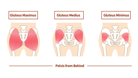
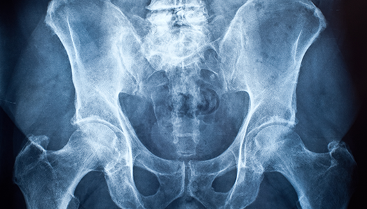
The iliopsoas (Psoas) is a muscle that originates within the pelvis and passes over the front of the hip joint before inserting on the femur (thighbone) at the lesser trochanter. As it travels from inside the pelvis to the femur it starts as a muscle and transitions to a tendon. The primary function of the iliopsoas is to flex the hip joint (serve to bring the knee to the chest). In some instances this tendon/muscle can become irritated and/or tight. Athletes and individuals who engage in sports or activities (such as running and sprinting) that involve rapid and repetitive hip flexion are at a greater risk for injury of this muscle/tendon. If the iliopsoas is tight for a long period, it may rub across the front of the hip joint and cause a snapping sensation and/or damage to structures of the hip, a scenario known as psoas impingement. Psoas impingment can be accompanied by significant pain and disability and when accompanied by a snapping sensation and/or sound is referred to as “internal snapping hip syndrome.” Dr. Anz will evaluate a patients symptoms and physical examination. The first step for treating psoas impingement is a trial of stretching, anti-inflammatories, physical therapy, and rest. In many cases, the inflammation will resolve and the symptoms decrease. A trial of physical therapy is necessary with a goal of guided stretching and hip strengthening. 
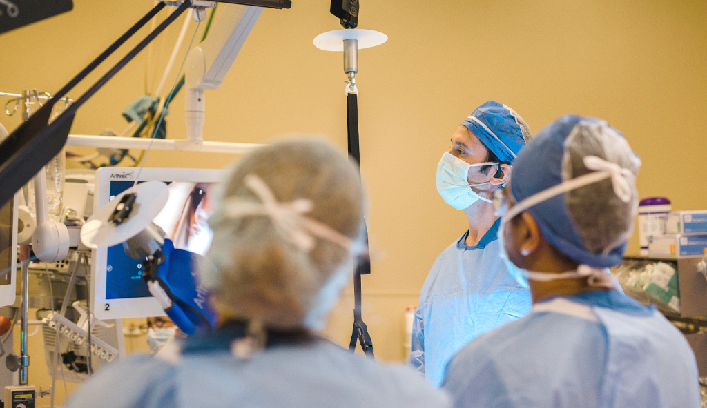
If pain and snapping sensations do not improve, Dr. Anz may recommend surgery to lengthen the muscle/tendon unit. The iliopsoas is an important stabilizer of the hip joint, so for some patients this may not be the best option. Dr. Anz will determine if this approach will offer the best outcome based on a patient-by-patient basis. When a psoas lengthening is deemed appropriate, he will use an arthroscopic approach. During surgery, keyhole incisions are used at the hip where he can insert a small camera and surgical tools to view the inside of the hip joint and perform the lengthening. To relieve tightness and eliminate the symptoms of the tendon rubbing over the front of the pelvis, Dr. Anz will release the tendon portion of the iliopsoas at the hip leaving the muscle portion of the iliopsoas intact. This lengthening leaves the hip flexor attached to the femur, but releases the tension creating the problem. Following arthroscopic hip surgery for psoas hip impingement, Dr. Anz will prescribe a through rehabilitation program. This will consist of a progressive plan that will involve week-by-week rehabilitation exercises and a set of recommended protocols. Depending on the length of prior symptoms and associated injury, most patients are able to return to their previous activities within 4-6 months following surgery. For additional information on the treatment for psoas impingement, or to learn more about arthroscopic hip surgery and partial psoas lengthening, please contact the Gulf Breeze, Florida orthopedic surgeon, Dr. Adam Anz located at the Andrews Institute. 
Hip Pain

Hip Labral Repair

Hip Pain

Injury Overview

The acetabular labrum is a type of cartilage that covers the rim of the hip socket (known as the acetabulum).  It may be injured as a result of a sports accident or an acute injury such as a hip dislocation or subluxation. It also can be injured gradually with time in settings of femeroacetabular impingment.  The labrum exists to provide stability to the hip joint mainly by creating a negative pressure seal at the hip, this aids smooth hip motion and flexibility.  Damage to the labrum may be a major source of hip pain and weakness for athletes. However, not all labral tears are associated with hip pain.  Some studies have shown that they may exist in patients without pain/dysfunction at the hip.

If a acetabular labral tear is identified, it is important to determine that it is associated with pain from the hip joint.  When a labral tear is identified, strengthening the muscles around the hip joint will improve function and decrease pain in most instances.  If initial strengthening measures do not improve a patient’s symptoms, Dr. Anz may recommend a hip labral repair using hip arthroscopy.  During this procedure, Dr. Anz will use a small camera and tiny instruments to enter the hip and view the acetabular labrum and surrounding structures. Treatment for the actual tear will involve shaving and smoothing out the torn portion of the labrum (debridement) and/or reattaching healthy segments with sutures (repair).  Repair involves sutures (strong string-like material) and absorbable anchors (strong screw-like implants) which have been developed specifically for the hip.  The sutures and anchors are used to secure the labral tissue to the base of the acetabulum.

In some situations the labral tissue may be damaged beyond repair.  This can occur in scenarios where femeroacetabular impingment has been present for many years.  In these instances, Dr. Anz may perform reconstruction of the labral tissue using a graft. The graft will come from the patient or from a cadaver in order to reconstruct the damaged labrum. The goal of labral repair or reconstruction surgery is to restore the negative pressure seal which the labrum creates, thus restoring near-normal biomechanics of the hip.

Following hip labral repair surgery, Dr. Anz will prescribe a week-by-week rehabilitation protocol to follow.  This will be a progressive process where the patient works with a therapist to regain full range of motion and strength back to the hip joint. Depending on the exact nature of the surgery and complexity of the injury, recovery is estimated at 4-6 months.

For additional information on labral tear repair surgery, or to learn more about arthroscopic hip surgery, please contact the Gulf Breeze, Florida orthopedic surgeon, Dr. Adam Anz located at the Andrews Institute. 

Gluteus Medius Injury

Gluteus Medius/Minimus Repair

Gluteus Medius Injury

The gluteus medius and gluteus minimus are muscles which are important for motion and stability of the hip joint.

 They are key to activities of daily living as well as sporting activities. The muscles originate on the pelvis and insert on the femur, at the greater trochanter. The greater trochanter is an area near the top portion of the femur (thighbone). In some instances the tendinous attachment of these muscles can undergo degeneration and/or detachment at their insertion site on the greater trochanter. This injury to the tendon can result in hip weakness, pain, and/or disability. Surgeons have recently begun utilizing arthroscopic surgery techniques for repair of these tendons where they insert on the greater trochanter.

In patients with hip pain and weakness, Dr. Anz will first evaluate for signs of tendon injury. If the gluteus muscles are found to be injured, some can improve with rest and therapy to strengthen muscles around the hip. Anti-inflammatory medication and stretching can also help decrease symptoms for some patients. If pain and functional limitations persist, surgery to repair the tendon at the attachment site may be helpful. The arthroscopic repair of the gluteus medius and/or minimus tendons is usually accompanied with removal of surrounding tissue that has become irritated, including inflammed greater trochanter bursal tissue. Additionally, a tight band of tissue overlying the area, the iliotibial band, may also be released. In some instances, can help decrease pain associated with inflammed bursal tissue, also know as bursitis.

During surgery, the torn gluteus medius or minimus tendon will be reattached to the native attachment site near the top of the femur on the greater trochanter. Suture anchors are embedded in the bone and strands of suture are then passed through the pulled-off portion of the tendon using specialized passing instruments. By tying these sutures, the torn tendon is once again attached to the bone.

Gluteus Maximus, Gluteus Medius, Gluteus Minimus

Following arthroscopic hip surgery for a gluteus medius repair or gluteus minimus repair, patients will need to avoid certain hip motions and exercises for 6 weeks. A brace helps to decrease stress at the repair site during this time as well.

For more information on arthroscopic hip surgery or for additional resources on the repair of a gluteus medius or minimus tear, please contact the Gulf Breeze, Florida orthopedic surgeon, Dr. Adam Anz located at the Andrews Institute.

Hip Runner Orthopedics

Chondroplasty

Hip Runner Orthopedics

Articular cartilage is a soft, fibrous tissue that covers the ends of bones within a joint, allowing for smooth, low-friction motion. Articular cartilage can become damaged through trauma associated with sporting accidents, femeroacetabular impingment, or as a result of the normal aging process. 

While certain structures within the hip joint may be capable of healing, articular cartilage has an extremely low healing potential. Chondroplasty is a procedure intended to remove mechanical symptoms related to cartilage injury and help prevent further injury to the cartilage. The goal is to delay the onset or progression of arthritis of the hip, which is often the result of cartilage damage.

When articular cartilage becomes damaged, loose flaps of cartilage may cause mechanical symptoms and/or can break off and become lodged in the hip joint. Eventually, cartilage damage may leave exposed bone resulting in bone-to-bone grinding sensations and a loss of joint motion. This is often associated with hip pain and hip stiffness, thus limiting movement and function. Arthroscopic hip chondroplasty is a procedure that Dr. Anz performs to remove mechanical symptoms and improve function. The approach has been shown to be most successful in patients with small, partial thickness injuries where the damage has not yet reached the bone itself and when the deforming force causing the cartilage injury can also be removed, such as in the setting of CAM femeroacetabular impingment. During this procedure, which is performed arthroscopically using tiny keyhole incisions, the loose flaps of cartilage are removed. Using a camera (arthroscope), Dr. Anz will be able to visualize where the loose pieces of fragmented cartilage are located, and will remove them. He will also address other abnormalities in the same surgery. In patients who are found to have exposed bone from loss of cartilage with good intact surrounding cartilage a microfracture procedure may also be performed to regenerative tissue. Although microfracture cannot restore normal cartilage, it does produce functional tissue.

Following arthroscopic hip chondroplasty, Dr. Anz will prescribe a thorough physical therapy and rehabilitation program. This will be a progressive process and will work on restoring motion, strength and full function of the hip joint. Patients who undergo a microfracture procedure can expect to be on crutches for 8 weeks.

Hip MRI

For more information on arthroscopic hip surgery, including chondroplasty of the hip for articular cartilage treatment, please contact the Gulf Breeze, Florida orthopedic surgeon, Dr. Adam Anz located at the Andrews Institute. 

A volleyball player jumps to spike the ball.

Hip Arthroscopy

A volleyball player jumps to spike the ball.

Dr. Adam Anz is trained in hip arthroscopy and uses this approach to treat a number of hip conditions.

Hip arthroscopy uses tiny, keyhole incisions to insert a small video camera (known as an arthroscope) and surgical tools inside the hip joint. The camera projects onto a high-definition video screen giving Dr. Anz and his surgical team a clear picture of structures in and around the hip.  This allows first-hand analysis of injury and can be done in order to repair and correct problems.

Historically, hip surgery has involved an open approach to the joint.  This involves making an incision on the skin and approaching the joint with a surgical dissection. Arthroscopic hip surgery has advanced in recent years making it possible to treat some problems with smaller incisions and without a surgical dissection.  While not all hip problems can be treated with hip arthroscopy, this technique opens the door for treatment of some hip injuries with a less invasive manner.

Dr. Anz uses an arthroscopic approach to address the following acute and degenerative conditions of the hip:

  • Femeroacetabular Impingment (also known as FAI Impingment)
  • Acetabular Labral Tears
  • Synovitis/PVNS
  • Psoas Tendon Pathology and Internal Snapping Hip Syndrome
  • Removal of Loose Bodies
  • Iliotibial Band Snapping and Trochanteric Bursitis
  • Articular Cartilage and Chondral Damage
  • Gluteus Medius and Minimus Tears
  • Hip Instability
Dr. Adam Anz performs surgery.

Following hip arthroscopy, it is important to follow the rehabilitation regimen that is set forth by Dr. Anz and by your physical therapist. Recovery following surgery is a partnership between the doctor, physical therapist, and patient. The results of the surgery are most effective when a post-operative rehabilitation program involving physical therapy and exercises are implemented daily. Dr. Anz can make recommendations for physical therapists in your area to help facilitate this process.

To learn more about hip arthroscopy, or to schedule a consultation please contact the Gulf Breeze, Florida orthopedic surgeon, Dr. Adam Anz located at the Andrews Institute. 

Acetabuloplasty

Acetabuloplasty (Pincer Impingement)

Acetabuloplasty

Femeroacetabular Impingment, often referred to as FAI for short, refers to a condition where there is a abnormality of the shape of the bones of the hip joint. 

FAI can include an abnormality of the femur, bone in the thigh, where it is shaped like a ball (Femoral Head), or FAI can include an abnormality of the pelvic bone where it is shaped like a socket (Acetabulum). In some scenarios, there may be abnormalties of both the femoral head and acetabulum. A pincer lesion is an anatomic abnormality of the acetabulum where there is too much bone covering the femoral head. The pincer lesion may cause impingement at the hip with certain hip motions. With time and repetition, impingement at the hip can lead to degeneration of the acetabular labrum and cartilage within the hip joint. A surgical treatment for pincer impingement is acetabuloplasty, where the bone causing the impingment is shaved with a burr.

Before Dr. Anz will recommend surgery for hip impingement, he will first try non-operative measures consisting of rest, activity modification, and physical therapy to strengthen the core musculature and muscles around the hip. Injections into the hip may also be used for diagnostic and therapeutic purposes. If a patient continues to have ongoing hip pain, acetabuloplasty surgery for hip impingement may be recommended. The goal of an acetabuloplasty is to correct the bony conflict by removing excess bone . Often this procedure will be coupled with measures to address any labral or cartilage damage that may also exist.

Dr. Adam Anz performs surgery at the Andrew's Institute.

During this procedure, Dr. Anz will use an arthroscopic surgical approach using two to three keyhole incisions. A camera (known as an arthroscope) is inserted into the hip to visualize the excess bone while the other incision is used to insert instruments to remove the excess bone. Often an arthroscopic burr is used to accomplish this. Hip acetabuloplasty entails taking away the area of over coverage on the acetabular side to relieve the bony impingement and create space allowing normal motion.

Following acetabuloplasty surgery for hip impingement, a thorough physical therapy program will be prescribed. This will include a progressive week-by-week plan that will work to restore motion of the hip while protecting repairs. Patients are typically on crutches for four weeks and placed in a brace to protect their hip during healing.

For more information on FAI impingement of the hip, pincer impingement of the hip, or for additional resources on treatments acetabuloplasty and arthroscopic hip surgery, please contact the office of Dr. Adam Anz, orthopedic hip surgeon in Gulf Breeze, FL.

A hockey player slides to defend a goal.

Fasciotomy (Iliotibial Band Release)

A hockey player slides to defend a goal.

The IT band (iliotibial band) is a thick band of fibers that runs the length of the outside of the hip and thigh—from the pelvis all the way down to the tibia (shinbone) just below the knee. 

Sometimes irritation and snapping can occur where this structure passes over a boney prominence known as the greater trochanter. This irritation is called bursitis and often occurs naturally as we get older. In athletes long episodes of training without rest or a proper stretching regimen can also cause bursitis and a tight IT band resulting in iliotibial band syndrome.

When patients present with IT band syndrome and/or greater trochanter bursitis, Dr. Anz will first recommend a trial of non-operative measures. This includes a period of rest, eliminating any activity that may flare the symptoms (e.g. running long distances), and methods aimed to decrease inflammation at the location. Physical therapy is often helpful to guide patients on methods to decrease the tension on the IT band as well as to help strengthen the adjacent musculature and core.

Surgery is treat IT band tension and bursitis is rarely necessary, and in some instances it may not be helpful. The rare instance where surgery is necessary is in cases of external snapping hip syndrome which does not respond to non-operative measures. Dr. Anz ultilizes an endoscopic technique to approach surgical cases of external snapping hip syndrome. First, he will perform an iliotibial band release (known as a fasciotomy). Using an arthroscope in an endoscopic fashion, Dr. Anz will get a visual of the IT band and will perform a small release in the fascia. 

Dr. Adam Anz performs a surgery at the Andrew's Institute.

He makes this release at the area of maximum tightness by cutting a diamond shape release at the area of the greater trochanter. The goal is to prevent further snapping sensations. This limited release also slightly lengthens the entire IT band. A second step is a trochanteric bursectomy. This involves the removal of inflamed bursal sac and irritated tissue.

Following surgery to treat IT band tightness, patients can be weight bearing as tolerated as long as an additional tendon repair has not been performed. Dr. Anz will prescribe a rehabilitation program that will focus on strengthening of core musculature and hip stabilizers in addition to continued IT band stretching.

For more information on IT band tightness, or to schedule a consultation to learn more about arthroscopic hip surgery, fasciotomy, or iliotibial band release, please contact the Gulf Breeze, Florida orthopedic surgeon, Dr. Adam Anz located at the Andrews Institute.

A soccer player rejoices with her team.

FCL Reconstruction

A soccer player rejoices with her team.

There are four major ligaments, or “restraints” to the knee joint.  They include the anterior cruciate ligament (ACL), posterior cruciate ligament (PCL), medial collateral ligament (MCL), and posterior lateral complex (PLC).  

The PLC is on the outer side of the knee and has three major components: the fibular collateral ligament (FCL, sometimes referred to as the LCL), the popliteus muscle and tendon, and the popliteofibular ligament.  The FCL can become damaged or torn with certain twisting injuries.  Commonly, this injury is sustained in high movement athletics such as football or soccer.  In some instances, the FCL may heal on its own after an injury; however, in certain instances, the ligament is torn to the extent that an FCL reconstruction is necessary.

By examining the knee and obtaining X-rays where the ligament is stressed, it is possible to determine which injuries will heal without surgery and which will require surgery.  If surgery is needed, Dr. Anz will perform an open FCL reconstruction, which will involve either harvesting one of a patient’s  hamstring tendons or the use of a donor tendon, which is also called an allograft.

After surgery, physical therapy is extremely important in order to obtain an optimal result.  Physical therapy begins immediately; however, knee range of motion is limited for 2 weeks.  Patients also cannot place weight on their leg for 6 weeks, as this is the amount of time which is necessary for tendon to heal to bone.  Strengthening begins at 8 weeks, and patients typically return to jogging at about 6 months depending on the extent of the injury.

Human Knee Diagram

For additional information on FCL knee injuries, or to learn more about FCL reconstruction surgery, please contact the Gulf Breeze, Florida orthopedic surgeon, Dr. Adam Anz located at the Andrews Institute.