Clavicle Fracture Fixation

Clavicle Fracture Fixation

Clavicle Fracture Fixation

The collarbone (clavicle) of the shoulder can become fractured from a direct hit or a fall onto the shoulder. This is a common injury for athletes and frequently occurs in football players.  Clavicle fractures can occur anywhere along the bone and in numerous configurations. They also can result in multiple bone fragments which is a scenario referred to as “comminution”.  Most clavicle fractures can heal over time with use of a sling and avoidance of activities for 6 to 10 weeks. In comminuted fractures or fractures in which the bones do not line up well, surgery is often recommended to restore the normal alignment and length of the bone.

Clavicle fracture surgery usually involves an incision over the top of the shoulder and placement of a plate and screws along the top of the clavicle. The goal of surgery is to stabilize the clavicle so that it can heal in the appropriate position. Surgery does not speed up the healing process but rather ensures that the bone heals correctly. Certain types of clavicle fractures can also be fixed with the use of a long pin placed within the bone (intramedullary nailing). Patients are usually allowed to go home the same day as their surgery.

Recovery from clavicle fracture surgery often takes a few months and involves sling use and guided physical therapy. This included range of motion training and strengthening. Some patients request to have their hardware removed from the collarbone but this is not allowed until after the fracture has fully healed.

For additional information on clavicle fracture fixation surgery, or to learn more about other injuries associated with the collarbone, please contact the Gulf Breeze, Florida orthopedic surgeon, Dr. Adam Anz located at the Andrews Institute. 

A patient exhibits rotator cuff pain.

Rotator Cuff Repair with Augmentation (Graft Reinforcement)

A patient exhibits rotator cuff pain.

The rotator cuff is made up of muscles and tendons that surround the shoulder joint. It is responsible for centering the ball in the socket of the shoulder and helps the shoulder to stay stable and move.   

Injuries to the rotator cuff are quite common. Sports are among the most common reasons why rotator cuff injuries occur. In addition, a traumatic occurrence such as a hard fall, or injury that occurs from micro-damage that stems from overuse and impingement (pinching of the tendon against the acromion process of the scapula) can also be factors. Chronic degenerative changes of these tendons can also make them weak and prone to tears. Injury to the rotator cuff may involve one or more tendons. The spectrum of injury can range from mild tendonitis, inflammation, and bursitis, to partial tears and full thickness tears.

In the case of a rotator cuff tear, surgery is usually required. In instances when the tear is acute and the tendon/muscle is healthy, the rotator cuff can usually be primarily reattached to the anatomic footprint using strong sutures and anchors placed in the bone. In unusual tear patterns, chronic retracted tears, degenerative tears, and tears that have some atrophy (loss of bulk) of the muscle, repairing the damage isn’t as easy.

In these situations, a technique referred to as rotator cuff repair with augmentation of graft reinforcement is usually used.  This is a safe and effective technique used to augment the repair providing better strength and a healing environment for the tendon.

Rotator Cuff Physical Therapy

This procedure is typically performed with a combination of an arthroscopic and open approach to the shoulder. The torn tendon is identified and a primary repair is attempted. At this point a decision is made as to whether the tendon would benefit from an augmentation. If this is the case a graft (patch) is obtained (which is made from cadaveric dermis).

The type of tear will dictate the exact configuration of the repair needed as well as the size of the patch. The patch is sewn into the rotator cuff tendon and a repair will involve using strong sutures and anchors into the bone to secure the graft and tendon.

In specific situations, adjuncts may be used to assist in healing such as a marrow stimulation healing response technique or an injection of platelet rich plasma.

Following rotator cuff repair surgery with augmentation, physical therapy becomes a crucial component in the recovery process. The specific progression of physical therapy following a surgery of this nature will depend on the configuration of the tear, type of repair that was used, and the number of tendons involved.  In most cases where a graft was used to help repair the injury, the progression of therapy is often slower.  It is very important that patients follow the advice of their therapists and take their time with this type of therapy so that the delicate graft and surgical processes are not interrupted. Therapy will typically consist of passive range of motion, followed by active motion, strengthening, and eventually return to activities. A sling is worn for 6-8 weeks after the surgery. The procedure has been shown to be highly successful in alleviating pain and improving function of the shoulder.

To learn more about the treatment alternatives for rotator cuff injuries, or to learn more about rotator cuff repair with a graft reinforcement augmentation, please contact the Gulf Breeze, Florida orthopedic surgeon, Dr. Adam Anz located at the Andrews Institute. 

A patient exhibits rotator cuff pain due to a rotator cuff injury.

Arthroscopic Rotator Cuff Repair

A patient exhibits rotator cuff pain due to a rotator cuff injury.

Rotator cuff injuries are common injuries among athletes. They occur when one of the four rotator cuff muscles are injured or damaged.   

The rotator cuff muscles involve four muscles that surround the shoulder and are responsible for stabilizing the ball in the socket of the shoulder. They work together to provide the shoulder with its powerful range of motion, strength, and the ability to perform overhead activities.

As the muscles get closer to their insertion on the humerus, they are called tendons. There are numerous activities that may cause damage to these tendons. Sometimes the damage is small and considered “micro” from overuse and impingement. Inflammation is very common condition as well. Other times, the injury may consist of a tear, or from chronic degenerative changes of the tendon. When injuries to the rotator cuff occur, a broad spectrum of symptoms can occur and treatment for the condition will depend on the type of injury or tear, and the number of tendons involved. 

In mild cases that may consist of small or partial tears, or tendonitis, Dr. Anz will usually try and treat the condition conservatively using non-operative measures first. These consist of rest, ice, anti-inflammatory medications, physical therapy, and possibly, an injection to reduce the inflammation.

In patients with full thickness tears, large partial-thickness tears, or smaller tears/tendonitis that have failed non-operative measures, surgery may be indicated. 

In the majority of cases, surgery to treat rotator cuff tears can be performed arthroscopically using small incisions, a camera, and tiny instruments to perform the procedure.

During surgery, Dr. Anz will enter the shoulder joint and examine the injury by identifying the torn tendon and reattaching it to its insertion site on the humerus—this is known as the footprint, or original site of attachment.  This process is completed using sutures placed through the tendon, and anchors that are attached to the bone to secure the normal anatomy of the tendon.

Rotator Cuff Xray

Dr. Anz will determine the exact type of repair once the configuration of the tear has been assessed. 

New procedures are now helpful in repairing tendons with additional support.  In some cases, a double row repair will be performed if needed to provide compression of the tendon for healing. Every patient will be given a strict rehabilitation protocol to follow once surgery is completed. Immediately following post-op, patients are placed into a sling and an individualized physical therapy program is started. Rehabilitation will include the work of a skilled therapist and will involve passive range of motion, followed by active motion, strengthening, and eventually return to activities. The specific progression of therapy will depend on the configuration of the tear, type of repair, and the number of tendons involved.

For additional resources on rotator cuff injuries, or to learn more about arthroscopic rotator cuff repair surgery, please contact the Gulf Breeze, Florida orthopedic shoulder practice of Dr. Adam Anz located at the Andrews Institute. 

Pole Vaulter sets her mark before beginning her vault.

Subacromial Impingement

Pole Vaulter sets her mark before beginning her vault.

Injury Overview

In many individuals, repetitive use of the shoulder can result in painful symptoms that make everyday activities difficult to perform. Subacromial impingement is one specific condition that affects adults, most commonly over the age of 30, and refers to the pinching (impingement) of the tendons of the rotator cuff between the humeral head and the acromion process of the shoulder blade. Irritation of the surrounding tendons leads to inflammation of the bursa which results in shoulder pain, and the inability to move the arm above the shoulder.  As the inflammation progresses, the symptoms become more severe and will occur more frequently.

In some cases, subacromial impingement may be associated with biceps tendonitis and/or rotator cuff tearing. Another factor that may cause shoulder impingement is weakness of the muscles around the shoulder blade, which cause it to sit in an inappropriate position and may predispose patients to the condition.  In addition, some patients have an abnormal shape to their bone, or have had a previous shoulder injury, both of which may be predisposed an individual to developing subacromial impingement.

Symptoms

The primary symptom associated with subacromial impingement is a sharp, piercing pain felt on the front of the shoulder with any type of overhead activity.  If the arm is resting, pain may not necessarily be felt, however, the arm will experience overall weakness making it difficult to use the affected arm for chores or activities.

Diagnosis

Dr. Anz will conduct a thorough physical examination that will include tests to check for pain level and mobility of the shoulder. Physical exam tests to assess for impingement include the impingement tests of Neer and Hawkins.  While shoulder impingement is relatively easy to diagnose based on the physical exam, Dr. Anz will require an X-ray and potentially a MRI to rule out if any other condition that may cause or contribute to the shoulder pain.

Treatment

In cases of subacromial impingement Dr. Anz will first recommend conservative treatment measures which consist of anti-inflammatories, rest, and physical therapy.  In many cases, these treatments are effective and surgery will not be needed. In cases with continued pain, an injection of corticosteroids to the subacromial bursa may help alleviate pain and allow the patient to make progress with physical therapy and accomplish longstanding relief.


Shoulder MRI

Surgical Treatment

In more serious cases of subacromial impingement, Dr. Anz may recommend a surgical treatment. Arthroscopic shoulder surgery is often used to correct this condition. This surgery is performed through several very small keyhole incisions through which a camera and special surgical instruments are inserted. During this operation, Dr. Anz will assess the rotator cuff to make sure there is no injury. The area of inflammation in the subacromial bursa is then removed. The bone spurs causing the pinching are also removed and the entire area is smoothed down to allow normal, pain-free motion. In complex cases of impingement, Dr. Anz may also treat other conditions that will present themselves once he is inside the shoulder joint.  These can also be addressed during the surgery, and may include arthritis between the clavicle and the acromion, as well as inflammation of the biceps tendon.

Post-Operative

Following surgery, Dr. Anz may request that the arm be kept in a shoulder sling for a period of time to allow healing.  Soon after, he will prescribe a thorough rehabilitation program. This program is a combined effort between the patient, Dr. Anz, and the physical therapist and will introduce exercises, strengthening moves, and active range of motion in time. It typically takes 2 to 4 months to achieve complete relief of pain, but in patients who presented complex cases, full recovery may take up to a full year.

For more information on subacromial impingement, or to discuss your shoulder pain and shoulder injury with Dr. Adam Anz, please contact the Gulf Breeze, Florida orthopedic surgeon, Dr. Adam Anz located at the Andrews Institute. 
Sternoclavicular

Sternoclavicular Joint Injuries

Sternoclavicular

The sternoclavicular (SC) joint is a major joint of the upper body that is formed by the articulation of the collarbone (clavicle) and the center of the chest (manubrium). 

Most of the SC joint’s strength and stability originates from the joint capsule and supporting ligaments. SC joint injuries are often due to a direct blow or blunt force trauma that occurs to the collarbone. These injuries can also result from a traumatic landing to the shoulder area (such as in football). SC joint injuries are typically associated with a disruption of the supporting ligaments.

A sternoclavicular (SC) joint injury is relatively uncommon, but when it occurs, the affected ligaments are stretched or torn (partially or completely) causing the joint to become disrupted. Sternoclavicular joint injuries are graded into 3 types ranging from a first-degree injury that involves a simple sprain or stretching of the ligaments, to a second-degree injury where a portion of the clavicle becomes subluxated. In the most severe cases (a third degree SC joint injury), a complete rupture typically occurs between the sternoclavicular and costoclavicular ligaments, which permits the clavicle to completely dislocate from the manubrium.

Symptoms

The most common symptoms from an SC joint injury include:

  • Severe pain at or around the SC joint at the time of injury and afterwards
  • Bruising in the area
  • Difficulty swallowing, difficulty breathing, a sense of fullness around the neck or a feeling of choking sensation due to posterior displacement of the medial clavicle
  • Cracking noises, popping sounds
  • A feeling of instability where the clavicle feels like it “moves” during activities


Dr. Adam Anz performs surgery at the Andrews Institute in Gulf Breeze, FL.

Diagnostic Testing

Dr. Anz will conduct a thorough examination of the shoulder blade and collarbone. He will test for tenderness and pain, and evaluate the overall range of motion of the arm and shoulder. In some cases, he will be able to see if a dislocation or other major disruption has occurred, but an X-ray will usually confirm his diagnosis. Because ligaments and other soft tissue structures are typically involved with these injuries, he may also order an MRI to take a more in-depth look at this area.

Treatment

Non-Surgical

Surgery is not necessary for most SC joint injuries. Initial treatment consists of ice, pain medication and sling immobilization. Return to activity is based on type of sport and the position played, the arm injured (dominant versus non-dominant), and the severity of the sprain. In more acute cases, a reduction (or relocation) of the SC joint can be attempted by sedating the patient and pulling gently on the arm, manipulating it back into its normal position. This may allow the SC joint to ‘pop’ back into position.

Surgical

Operative treatment is reserved for patients who experience major trauma to the bones and ligaments of the SC joint. Dr. Anz will choose the type of surgery based on the exact injury, but in many cases will opt for a stabilization procedure that will allow him to reattach or repair any loose or torn ligaments of the SC joint. Without this stabilization procedure, a patient is a risk for future dislocations and/or subluxations, as well as the onset of arthritis.

Post-Op Recovery

Following surgery for an SC joint injury, it is critical that patients follow the prescribed post-op rehabilitation program as set forth by Dr. Anz. These guidelines will be broken down into phases and will be conducted with a therapist, and at home. Most patients are able to resume normal activities within 3-6 months; sports and other high impact activities may take longer.

For more information on sternoclavicular joint injuries or for additional resources on the other shoulder related injuries and conditions, please contact the Gulf Breeze, Florida orthopedic surgeon, Dr. Adam Anz located at the Andrews Institute. 
Family Hiking Outdoors

Shoulder Fractures

Family Hiking Outdoors
Fractures that occur within the structure of the shoulder joint can appear as a break or a crack in the bone on an X-ray. A shoulder fracture is generally classified based on which bone (clavicle, humerus, or scapula) is involved. A shoulder fracture can occur when sudden force, impact, or trauma has been sustained to the shoulder joint. Hard falls during sporting events or motor vehicle trauma are examples of possible causes of fractures in the shoulder. There are 3 distinct bones within the shoulder that could sustain a fracture: The collarbone (clavicle) is the most common place where a fracture may exist; the upper arm bone (proximal humerus) can be fractured and is often related to poor bone density; a fracture of the shoulder blade (scapula) is less common, but usually occurs during a high-energy impact. Fractures are classified as being displaced or non-displaced. In a non-displaced fracture, the broken pieces line up on each side of the break. Displaced fractures, in which the pieces on either side of the break are out of line, may require some type of manipulation to restore normal anatomy. Occasionally the rotator cuff muscles are injured or torn at the same time as the fracture, which can further complicate the treatment.

Symptoms

Shoulder fractures are usually characterized by pain with motion or palpation of the shoulder. Other symptoms include swelling or bruising of the shoulder area, a bump or bulge at the site of the break, or an inability to move the arm without pain.

Diagnostic Testing

A physical examination reveals pain over the bone, and often swelling or bruising is present. X-rays typically show the fracture. However, in more serious cases, 3-D imaging, such as CT scans or MRIs are often ordered to get a better picture of the fracture pattern.

Treatment

Non-Surgical Many fractures can be treated without surgery, and heal in about 4 months. Many shoulder fractures can be treated with a simple sling and limited use of the shoulder until the pain and inflammation has subsided. By immobilizing the shoulder, the bone is allowed to heal, and once it has healed enough, physical therapy is typically ordered to regain motion and strength. Surgical The need to surgically repair a shoulder fracture depends upon your age, activity level, and the severity of your injury. Some fractures are better treated with surgery because they may carry a high risk of arthritis if left alone. There are many different types of surgery for different types of fractures including percutaneous pinning (placing pins in the bone to secure them back in place); open reduction internal fixation (ORIF—which involves opening up the fracture, realigning the bones, and putting a plate and screws in place to hold them together); and shoulder hemiarthroplasty – replacing the shoulder if the fractures are in too many pieces.

Post-Op

After any fracture surgery, proper rest and rehabilitation is critical to good success and improvement of pain and function. Dr. Anz will prescribe a very detailed physical therapy program that, if performed consistently and successfully, will help rehabilitate the injured arm. Most patients can resume normal activities within six months following surgery. For additional information on shoulder fractures or to learn more about treatment for this shoulder condition, please contact the Gulf Breeze, Florida orthopedic surgeon, Dr. Adam Anz located at the Andrews Institute. 
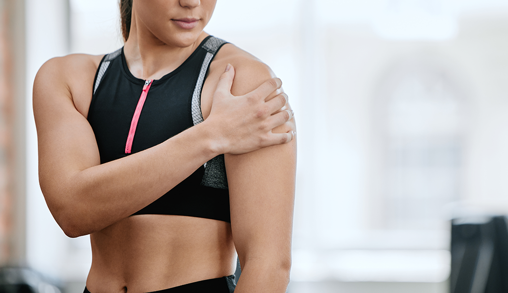
Scapulothoracic Bursitis

Scapulothoracic Bursitis (Snapping Scapula)

Scapulothoracic Bursitis

Injury Overview

The scapulothoracic joint is located in an area where the shoulder blade (scapula) meets and moves along the chest wall. The motion of the scapula is normally smooth because of the underlying scapulothoracic bursa, a fluid filled sac that aids in the movement of the scapula against the ribcage. If the bursa becomes inflamed from injury or overuse, a condition known as scapulothoracic bursitis may develop.

Scapulothoracic bursitis, also referred to as snapping scapula syndrome, is most often seen in young athletes who participate in repetitive overhead activities. However, the condition may develop in patients of any age. This syndrome is fairly rare and can also occur in individuals who sit or stand in a abnormal position for long periods of time and/or in patients whose shoulder movement was altered for long periods of time, such as in the setting of a shoulder injury.

Symptoms

Symptoms associated with a snapping scapula include pain located near the middle of the upper back (the top inside border of the shoulder blade) with movement of the shoulder. During shoulder movement, snapping, grating, and/or grinding noise is present. This is often accompanied by a dull ache in the shoulder blade and ribcage area. These symptoms may steadily progress with time.

Diagnosis

The most common physical exam finding in patients with this problem includes a grinding or snapping sensation over the inner aspect of the shoulder blade when the arm is moved. Dr. Anz will perform movement tests involving the shoulder joint to assess these symptoms. If he suspects that scapulothoracic bursitis is the condition causing the symptoms, he will most likely require an X-ray and MRI to confirm his diagnosis and to rule out any alternative abnormality. Once snapping scapula has been diagnosed, Dr. Anz will begin discussing treatments options according to the severity of the injury.

Treatment

Dr. Anz will prescribe a course of physical therapy to improve shoulder kinetics as the first line treatment for this condition. To manage pain during this recovery process, he may use a corticosteroid injection to the scapulothoracic bursa to relieve pain and allow further progress with physical therapy. Rehabilitation will focus on restoring normal scapular motion, eliminating positions which cause symptoms, and strengthening the musculature around the shoulder blade.

Surgical Treatments

If conservative treatments fail to ease the symptoms of snapping scapula syndrome and restore shoulder function, Dr. Anz may recommend surgery. In most cases, a minimally invasive arthroscopic approach can be used. This involves tiny keyhole incisions, a miniature camera, and surgical tools to assess and treat the bursitis. During the surgery, Dr. Anz will remove the inflamed scapulothoracic bursa, as well as areas of bone which may be attributing to the bursitis. These procedures are typically successful in relieving pain, eradicating symptoms of rubbing and snapping, and removing the areas of inflammation to restore shoulder motion.

Post-Operative

Dr. Anz will require patients to adhere to a strict rehabilitation program following arthroscopic surgery. Detailed guidelines, rehabilitation progression, and expectations for each of his patients will be outlined so that a full recovery can be achieved. Therapy is a vital part of the recovery process following a shoulder injury and is a partnership between the patient, Dr. Anz, and the physical therapy team.

For more information on snapping scapula syndrome or for additional resources on shoulder pain or scapulothoracic bursitis symptoms, please contact the Gulf Breeze, Florida orthopedic surgeon, Dr. Adam Anz located at the Andrews Institute. 
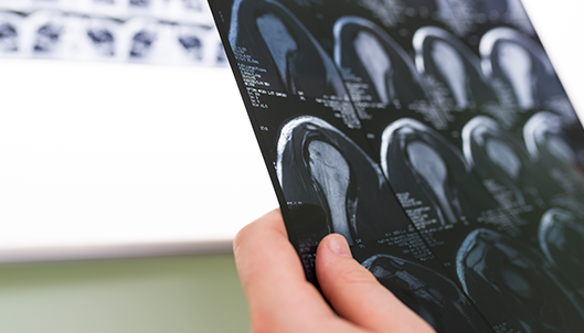
Rotator Cuff Injury

Rotator Cuff Injuries

Rotator Cuff Injury

Injury Overview

Rotator cuff injuries are a very common cause of shoulder dysfunction in both young and old patients. The rotator cuff is comprised of a group of four muscle-tendon units that surround the shoulder joint and allow for overhead motion of the arm. The muscles and tendons that make up the rotator cuff also help stabilize the shoulder joint. A rotator cuff injury can occur when any of these muscle-tendon units become damaged. This is typically characterized by a separation or “tearing” of the tendon attachment off of the humerus. This can occur due to a number of factors, including:

  • Acute injury – Such as a hard hit or traumatic fall sustained in a sport or car accident
  • Chronic overuse – Continuous, repetitive movement that occurs with athletic training in activities such as baseball (pitching) or performing overhead movements like in tennis or swimming. On the job lifting of heavy objects can also lead to overuse.
  • Gradual aging – the degeneration of the muscles or tendons over time and normal wear and tear can cause a breakdown of collagen, thus making the tendons and muscles more prone to degeneration and injury.

When any of these risk factors are present, the rotator cuff may be at risk for a tear. Certain other conditions, such as impingement and tendonitis, can also contribute to tearing of the rotator cuff. These tears result in a significant amount of shoulder pain, weakness, and limited range of motion. The severity of the injury may range from a mild strain with associated inflammation to a partial or complete tear that might require surgery.

Symptoms

The symptoms of a rotator cuff injury arise from the inflammation that accompanies the structural damage. The most common symptom is pain over the top of the shoulder and arm. In some patients, the pain can descend down the arm towards the elbow. This pain is evident at rest and may interfere with sleep at night, especially when lying on the affected shoulder.

Another common symptom typically associated with a rotator cuff injury includes weakness of the shoulder in combination with pain, causing difficulty in lifting the arm up in overhead motions or performing seemingly simple tasks such as getting dressed or reaching up to grasp an object.

 Diagnostic Testing

Dr. Anz will conduct a thorough examination and will observe the shoulder joint to look for deformities, muscle wasting, or weakness of the affected rotator cuff muscles. He will touch and manipulate the bones and muscles that make up the shoulder joint to find areas of tenderness or pain. He will carefully check overall mobility and strength. In addition, a variety of tests may be utilized to determine which of the muscles or tendons of the rotator cuff is damaged including X-rays (to look at the joint) or an MRI (to assess the soft tissue and cartilage).

Treatment

Most rotator cuff injuries can initially be treated without surgery. The success of non-surgical treatment options depends on the type of rotator cuff injury (acute versus more established), the size of the tear as determined by the diagnostic tests, and the activities of the patient.

Non-surgical

The goal of treatment is to reduce inflammation and strengthen the uninjured muscles around the shoulder to compensate for the torn or injured muscles or tendons. Some conditions may be easily treated with a combination of ice and heat, rest and diminished use of the shoulder for a determined amount of time. Anti-inflammatory medications (NSAIDs) can also help.

Some patients may require the assistance of a physical therapist to help strengthen the muscle and increase flexibility through a special exercise program. Dr. Anz may inject a steroid, such as cortisone, into the area of inflammation if the pain persists. The cortisone shot is designed to act only in the area where it is injected and has a potent anti-inflammatory effect.

Surgical

The need to surgically repair a rotator cuff injury depends upon your age, activity level, and the severity of your injury. Dr. Anz strives to achieve maximum recovery with minimal surgery and often prefers an arthroscopic treatment approach:

Arthroscopic Rotator Cuff Repair

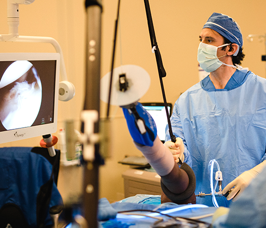
An arthroscopic rotator cuff repair is done through a series of small incisions. Dr. Anz utilizes a small camera to view the damaged tissue on a large television screen. He is then able to insert small instruments into the joint to achieve the repair. The advantage of arthroscopic surgery is that there is usually less pain and a faster return to normal activities.

Post-Op

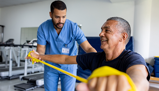
A rehabilitation program will be prescribed at your first post-operative visit with Dr. Anz. This will usually entail a period of rest and minimal rehab to allow the repair to heal. After healing is achieved, a more aggressive regimen is prescribed with the goal of regaining motion and strength.

Rehabilitation after surgery is as important as the surgical repair itself. Without proper rehab, the chance of full recovery is diminished. Patients are advised to be good to the shoulder as the rotator cuff heals by avoiding extra and unnecessary physical stress.

For more information on rotator cuff injuries, or for additional resources on shoulder injuries, please contact the Gulf Breeze, Florida orthopedic surgeon, Dr. Adam Anz located at the Andrews Institute. 

Labral & Slap Tear

Labral and SLAP Tears

Labral & Slap Tear

Injury Overview

The labrum is a type of cartilage found in the shoulder that surrounds the socket (glenoid) and has two primary functions: 1) to deepen the socket so that the ball of the shoulder stays in place; and 2) the Labrum acts as an attachment site for other structures, such as the biceps tendon or shoulder ligaments. When there is an injury to the shoulder, such as a dislocation, the labrum can be peeled off of the rim of the socket (glenoid).

Labral tears can occur for a variety of reasons and there is a number of ways that this injury can affect a patient. The most serious is when the labrum is torn completely away from the bone. This acute, traumatic injury is often associated with a dislocation or subluxation of the shoulder. Another labrum injury is associated with a tear within the substance of the labrum itself. When this degenerative condition occurs, the labrum is left with an unsmooth, rough edge. This condition is usually found in older patients. A tear can also occur in the area where the biceps tendon attaches to the upper end of the socket (SLAP tear).

A SLAP Tear (Superior Labrum Anterior Posterior) refers to a specific type of labral tear in the shoulder, which is located at the top of the shoulder socket (glenoid) and involves the attachment site of the biceps tendon. Acute trauma and overuse are often the causes of this specific type of labrum injury.

Symptoms

A SLAP tear is often accompanied by pain deep in the shoulder, stiffness, a popping or clicking sensation or feeling of instability. Decreased range of motion or strength can also be present.

Diagnostic Testing

Dr. Anz will conduct a thorough examination and perform a physical examination to help determine whether or not you have a SLAP tear. During the evaluation, Dr. Anz will determine if the tear is associated with any type of pre-existing instability to the shoulder or if it’s from a particular traumatic event. X-rays will rule out any fractures or bone-related issues. Typically, an MRI is most effective in diagnosing a tear.

Treatment

Several key factors play a role in the decision-making process regarding treatment of a tear, including age, type of tear, and athletic profile.

Non-surgical

Initially, patients are directed to rest and ice the area, along with taking anti-inflammatory medications followed by a course of physical therapy.

Surgical

When non-surgical treatments fail, arthroscopic surgery of the shoulder is typically recommended. Generally speaking, there are three surgical options for a SLAP tear

  • Debridement: During a debridement procedure, Dr. Padelecki will smooth out the torn labrum during an arthroscopic surgical approach. This option is only suitable for stable SLAP lesions that do not seem to involve the biceps tendon.
  •  SLAP Repair: A SLAP repair is an arthroscopic procedure that uses anchors with sutures attached to secure the torn labrum down to the shoulder socket. A SLAP repair is the most common procedure done for symptomatic SLAP lesions and is typically reserved for young patients with an otherwise healthy shoulder who want to remain athletically active.
  • Biceps tenodesis: A biceps tenodesis cuts the biceps tendon from where it attaches to the labrum, and reattaches it to another area. By decreasing the forces that pull on the SLAP region, the symptoms will be alleviated. A biceps tenodesis can either be performed arthroscopically, or through a small incision.

Post-Op

A rehabilitation program will be prescribed at your first post-operative visit with Dr. Anz. Rehabilitation after surgery is extremely important, especially in athletic individuals who are seeking to get back into regular activity. Without proper rehab, the chance of full recovery is diminished and shoulder stiffness can occur.

For additional information on labral and slap tears, or to learn more about arthroscopic shoulder surgery, please contact the Gulf Breeze, Florida orthopedic surgeon, Dr. Adam Anz located at the Andrews Institute. 
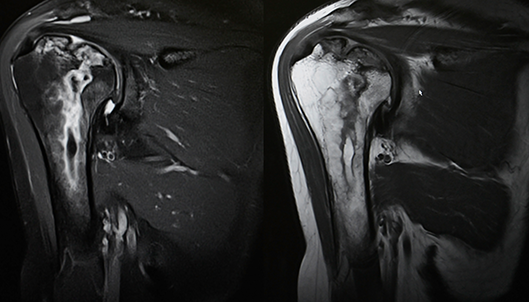
Shoulder Pain

Frozen Shoulder

Shoulder Pain
Frozen shoulder, also known as adhesive capsulitis, is a condition characterized by inflammation and stiffness in the shoulder joint which can restrict motion and cause chronic pain. Also referred to as “stiff shoulder”, this condition develops when the soft tissue of the shoulder joint begins to thicken and contract as a result of the formation of scar tissue. The scar tissue (referred to as “adhesions”) creates the loss of motion. The condition usually comes on slowly, but generally worsens over time. With the right treatment, frozen shoulder usually can be corrected and the symptoms alleviated.

Symptoms

The most common symptom of frozen shoulder is pain, which is typically described as dull or aching. It is worse early in the course of the disease and is usually located over the outer shoulder area and sometimes the upper arm. Limited mobility, shoulder weakness, and loss of movement of the joint are also major symptoms associated with frozen shoulder.

Diagnostic Testing

Dr. Anz may suspect frozen shoulder during a routine examination and after questioning of your past medical history. An X-ray or an MRI may be performed to see whether symptoms are from another condition such as arthritis, rotator cuff tear, or a broken bone.

Treatment

Non-Surgical Treatment for frozen shoulder usually starts with basic activities including rest, ice, and medication. Physical therapy is often very effective in treating frozen shoulder, especially when combined with steroid injection. These treatments should help the return of motion and function to the shoulder. Surgical If conservative treatments are not curing the frozen shoulder over a period of time, Dr. Anz will perform arthroscopic shoulder surgery to repair the condition. Prior to surgery, he may attempt a manipulation procedure under anesthesia, where he will force the shoulder joint to move, causing the capsule and scar tissue to stretch or tear. This releases the adhesions and increases range of motion. During arthroscopic surgery, Dr. Anz will attempt to cut some of the tight tissues around the shoulder and relieve the pressure.

Post-Op

Following arthroscopic shoulder surgery, patients will need to adhere to a rehabilitation program as instructed by Dr. Anz. This will involve specific exercises and movements over a distinct period of time. It’s in the patient’s best interest to complete rehab and work with Dr. Anz towards a complete recovery. For additional resources on available treatment for frozen shoulder, please contact the Gulf Breeze, Florida orthopedic surgeon, Dr. Adam Anz located at the Andrews Institute.